I was personally shocked by these photos and article.
Lennart Nilsson is a Swedish photographer and scientist, a pioneer of medical photography. He was born in 1922 and from childhood became interested in that part of the world around him that can only be seen through a microscope. When Lennart was 12 years old, his father, who was a photographer, gave him his first camera. Subsequently, the microscope and camera will become Nilsson’s main instruments, which will open to society fascinating pictures of the cosmic expanses of the human body and other spaces inaccessible to vision.
In the mid-1960s, ultra-thin endoscopes became available to science, thanks to which Nilsson began to take his pioneering images of life in the human body at the cellular level.
Nilsson gained international fame in 1965, when LIFE magazine published 16 pages of photographs of a human embryo. These photographs were immediately reprinted in Stern, Paris Match, The Sunday Times and other magazines.
Nilsson became a member of the Swedish Society of Medicine in 1969, and also received an honorary doctorate in medicine from the Karolinska Institutet in 1976. He continues his scientific and photographic activities to this day.
So, how does life begin:
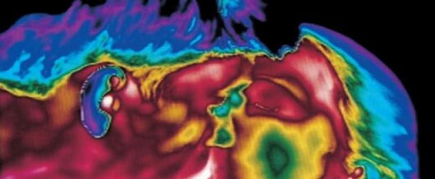
A photo of a kiss that reflects body temperature.

Millions of sperm swim towards the cervix.

Sperm in the fallopian tube.

Will the meeting take place?

Two sperm come into contact with the shell of the egg. Enzymes contained in the head of the sperm dissolve the membrane of the egg, but only the genetic material of one sperm is involved in fertilization.

One of the father's 200 million spermatozoa, having broken through the membrane of the egg, literally pours into it...

Longitudinal section of a spermatozoon. The genetic material is contained in the head of the sperm.

After a week, the embryo, sliding down the fallopian tube, moves into the uterus...

Embryo attached to the uterine lining (+8 days).

Embryo development. Gray color - future brain. (+22 days).

24 days. A one-month-old embryo does not yet have a skeleton - there is only a heart, it begins to pulsate on the 18th day.

28th day after fertilization...


4 and a half weeks...

A five-week embryo, 9 mm long, can already discern a face with holes for the mouth, nostrils and eyes.


40 days. The outer cells of the embryo grow together with the loose surface of the uterus and form the placenta, or baby's place. This spongy piece of flesh serves a person in the first nine months of his life as lungs, stomach, liver, and kidney...

Eight weeks. The rapidly growing embryo is well protected in the mother's womb. Using an electron microscope, Nilsson was able to magnify the image hundreds of thousands of times.

10 weeks. The eyelids are already half open. They will be fully formed within a few days.


Heart cells

Embryo leg

Fetal handle

16 weeks. The inquisitive baby is already using his hands to explore his surroundings.

The skeleton consists mainly of a flexible shaft and a network of blood vessels visible through thin skin.

18 weeks. About 14 cm. The embryo can now perceive sounds from the outside world.

20 weeks. About 20 cm. Hair is already beginning to appear on the head.

24 weeks...

26 weeks...

6 months. There are still eight to ten carefree weeks ahead, but the little man is already cramped in the uterus, and he is preparing to leave it. He turns upside down - it’s easier to get out...

36 weeks. After 4 weeks the baby will see the white light.

Today, August 24, the Swedish photographer and scientist, the man who went down in the history of photography with photographs of human embryos taken in natural conditions, the first modern photojournalist in Switzerland, celebrates his 93rd birthday. Lennart Nilsson.
Lennart Nilsson was born into a family of photographers, his father and uncle were photographers. When Lennart was 12 years old, his father gave him his first camera. At 15, Lennart watches a documentary about Louis Pasteur and becomes fascinated by microscopy. Over the next few years, Lennart Nilsson acquired a microscope and took microscopic photographs of insects.
His professional career as a photographer began in the mid-40s. He became a freelance photographer for the Stockholm publication Ahlen & Akerlund. One of Lennart's first assignments was to create a series of photographs dedicated to the liberation of Norway in 1945. Some of the photographer's early works, published in Life magazine, such as "Midwife in Lapland", 1945, "Hunting on Spitsbergen", 1947, "Fishermen on the Congo River", 1948, brought the photographer international popularity.
In 1954, 87 portraits of famous Swedes by Lennart Nilsson were published in the large publication Sweden in Profile. In 1955, the photographer published his own book called “Report,” which included the author’s early works. In 1963, the book Hallelujah, dedicated to the Swedish Salvation Army, was published.

In the mid-50s, Lennart Nilsson began experimenting with new photographic techniques, trying to get the maximum possible close-up in photography. His personal achievements in this, magnified by the thin and sensitive endoscope that became available by the mid-60s, allowed the photographer to create pioneering photographs of human blood vessels and the insides of the body at the highest possible magnification. In 1965, Lennart Nilsson achieves worldwide popularity thanks to the fact that a photograph depicting a human embryo at an early stage of development appears on the cover of Life magazine. Lennart Nilsson's photographs of human embryos travel from publication to publication. In 1965, a book entitled “The Birth of a Child” was published.

No matter how inconvenient and immoral it may be, the fact remains that in order to show the development of a living fetus, Lennart Nilsson used aborted material and photographed the fetus after the abortion, i.e., already dead. Working with dead material allowed the photographer to conduct more successful experiments with lighting, backgrounds and compositions. However, the origins of the photographs and the photographer's working methods are rarely mentioned.
In 1969, the photographer began using a scanning electron microscope to obtain images of the internal functions of the human body. Lennart Nilsson is credited with taking the first photograph of the human immunodeficiency virus, and he also became the first photographer to take a picture of the SARS virus in 2003.


Nilsson gained international fame in 1965 when LIFE magazine published 16 pages of photographs of a human embryo. These photographs were also immediately reproduced in Stern, Paris Match, The Sunday Times and other magazines, bringing photographer Lennart Nilsson worldwide fame.
Since childhood, the microscope and camera were the main hobbies of Nilsson, who wanted to show the whole world the beauty of the human body at the cellular level. Nilsson managed to obtain photographs of the human embryo back in 1957, but they were not yet impressive enough to be shown to the general public.
A cystoscope, a medical device used to examine the inside of the bladder, helped him get the most accurate and colorful images. Nilsson attached a camera and a light guide to it and took thousands of pictures of the baby's life inside the uterus.
Thus, the skillful hands of Lennart Nilsson created a miracle: they showed the whole world the mystery of the origin of human life.
The sperm in the fallopian tube moves towards the egg
 Egg
Egg
 Fateful meeting
 One of the father's 200 million sperm breaks through the egg membrane
 Spermatozoon in section. The head contains all the genetic material.
 A week later, the embryo, sliding down the fallopian tube, moves into the uterus
 After another week, the embryo attaches to the lining of the uterus
 Day 22 of embryo development. Gray matter is the future brain
 On the 18th day, the embryo's heart begins to pulsate
 28th day after fertilization
 5 weeks, length 9 mm, a face with holes for the mouth, nostrils and eyes can already be guessed
 40 days. The outer cells of the embryo grow together with the loose surface of the uterus and form the placenta
 10 weeks. The eyelids are already half open. They will be fully formed within a few days.
 10 weeks. The baby is already using his hands to explore his surroundings
 16 weeks
 A network of blood vessels is visible through thin skin
 18 weeks. The embryo can perceive sounds from the outside world
 19 weeks
 20 weeks. Height is about 20 cm. Hair begins to appear on the head
 24 weeks
 6 months
 36 weeks. In a month the baby will be born.
In 1965, a book of Nilsson's photographs, A Child is Born, was published and sold out within a few days. The book has gone through many reprints and still remains one of the best-selling books in the history of this kind of photo albums.
Medical photography pioneer Lennart Nilsson is now 92 years old. He is still involved in science and photography.
Swedish photographer Lennart Nilsson showed the world photographs of the origin of human life, from conception to birth.
The world heard about Lennart Nilsson back in 1965, when his photos were published on the pages of LIFE, which depicted a human embryo at all stages of its development. The photographs immediately spread across various publications.
Microscopes and cameras have been Nilsson's passion since childhood. Over time, ambitions formed into a profession to show the world the beauty of the birth of human life from the very beginning. He managed to take the first photographs of the fetus already in 1957, but their quality left much to be desired.
Nilsson managed to get the perfect shots using a medical tool for examining the bladder - a cystoscope, to which was attached a camera with a tiny light source. It was with the help of this device that photographs were taken recording the life of the embryo in the womb.
Nilsson created something truly miraculous: for the first time, people could see with their own eyes the conception and early development of human life.
Lennart Nilsson died on January 28, 2017 at the age of 95. Until the end of his days, he never ceased to be interested in science and photography.
20 PHOTOS
1. The sperm moves down the fallopian tube towards the egg to fertilize it.  2. Photo of the egg.
  3. Decisive moment.
  4. Out of hundreds of millions of sperm, only one can fertilize an egg.
  5. The genetic material is located in the head of the sperm.
  6. After a week, the embryo begins its journey to the uterus to attach to its walls.
  7. In another week, the embryo will attach to the wall of the uterus.
  8. Embryo at 22 days gestation. The gray area becomes the child's brain.
  9. By the 18th day, the fetus’s heart begins to beat. If you have been putting off making an appointment with a gynecologist until now, now is the time to do it. The gynecologist will prescribe an ultrasound and determine that the fetus is normally attached to the wall of the uterus.
  10. 4 weeks after fertilization.
  11. At five weeks, the fetus is 9 millimeters long. The photo shows a developing face, and the holes are the future nostrils, mouth and eyes.
  12. 6 weeks of development. The outer cells of the embryo join the free surface of the uterine wall, forming the placenta, through which the fetus receives all its nutrients.
  13. 8 weeks after conception.
  14. 10 weeks after conception. The eyelids are half open. In a few days they will be fully formed.
  15. After 10 weeks, the embryo uses its arms to explore the world around it.
  16. 16 weeks after conception.
  17. Blood vessels are visible through the skin.
  18. 18 weeks. The fetus can now hear sounds from the outside world.
Not for children about children or life before birth Many photographs are the work of Swedish photographer Lennart Nilsson. In 1957, Lennart Nilsson began taking photographs using an endoscope, an instrument that allows one to see the inner world of a person. When he presented his work to the editors of Life magazine, they did not believe him at first. Now an hour of photographs are the result of computer processing, but they do not lose their value because of this. The technique of medical photography brought him worldwide fame. The sperm in the folds of the mucous membrane of the fallopian tubes moves towards the egg.  Longitudinal section of a spermatozoon. The genetic material is contained in the head of the sperm.
  Before entering the cervix, sperm represent the 500 millionth cohort at the start! When approaching the germ cell, there are no more than a hundred of them, capable of destroying its protective shell.
  From 3-7 hours after ejaculation: sperm “unwind” the egg and only the strongest and luckiest sperm will participate in fertilization.
  Embryo attached to the uterine lining (8 days)
  20 hours after ejaculation: Inside the fertilized egg, the nuclei of the male and female cells unite and new chromosomes (genetic material) are formed.
  1st day after fertilization: the cell begins its journey from the tube where fertilization occurs to the uterus. Two days after the meeting of the “winners”: four new cells, surrounded by small nutrient “communities”, glide towards the uterus.
  4th day. Morula stage.
  Embryo development. Gray color - future brain. (22 days)
  The heart is formed and begins to beat. (24 days)
  28 days pregnant
 40 days of pregnancy: placenta, umbilical cord and embryo can be seen Fourth week of life. The size of the embryo is 6 mm. You can already discern the outlines of the back and brain. A convex ball is visible - the yolk sac, a small embryonic appendage that will disappear by 11
- In contact with 0
- Google+ 0
- OK 0
- Facebook 0