Viruses are microorganisms that make up the kingdom Vira.
Features:
2) do not have their own protein-synthesizing and energy systems;
3) do not have a cellular organization;
4) have a disjunctive (separated) method of reproduction (synthesis of proteins and nucleic acids occurs in different places and at different times);
6) viruses pass through bacterial filters.
Viruses can exist in two forms: extracellular (virion) and intracellular (virus).
The shape of virions can be:
1) round;
2) rod-shaped;
3) in the form of regular polygons;
4) thread-like, etc.
Their sizes range from 15–18 to 300–400 nm.
In the center of the virion is a viral nucleic acid, covered with a protein shell - a capsid, which has a strictly ordered structure. The capsid shell is made up of capsomeres. Nucleic acid and the capsid shell make up the nucleocapsid.
The nucleocapsid of complexly organized virions is covered with an outer shell - a supercapsid, which can include many functionally different lipid, protein, and carbohydrate structures.
The structure of DNA and RNA viruses is not fundamentally different from the NK of other microorganisms. Some viruses contain uracil in their DNA.
DNA can be:
1) double-stranded;
2) single-chain;
3) ring;
4) double-stranded, but with one shorter chain;
5) double-chain, but with one continuous and the other fragmented chains.
RNA can be:
1) single thread;
2) linear double-stranded;
3) linear fragmented;
4) ring;
Viral proteins are divided into:
1) genomic – nucleoproteins. Provide replication of viral nucleic acids and viral reproduction processes. These are enzymes, due to which the number of copies of the parent molecule increases, or proteins, with the help of which molecules are synthesized on a nucleic acid matrix that ensure the implementation of genetic information;
2) capsid shell proteins are simple proteins with the ability to self-assemble. They form geometrically regular structures, in which several types of symmetry are distinguished: spiral, cubic (they form regular polygons, the number of faces is strictly constant) or mixed;
3) supercapsid shell proteins are complex proteins with diverse functions. Due to them, the interaction of viruses with a sensitive cell occurs. Perform protective and receptor functions.
Among the proteins of the supercapsid shell are:
a) anchor proteins (one end of them is located on the surface, and the other goes deep; they ensure contact of the virion with the cell);
b) enzymes (can destroy membranes);
c) hemagglutinins (cause hemagglutination);
d) elements of the host cell.
2. Interaction of viruses with the host cell
The interaction takes place in a single biological system at the genetic level.
There are four types of interaction:
1) productive viral infection (interaction as a result of which virus reproduction occurs and cells die);
2) abortive viral infection (an interaction in which virus reproduction does not occur, and the cell restores the impaired function);
3) latent viral infection (the virus reproduces, but the cell retains its functional activity);
4) virus-induced transformation (an interaction in which a cell infected with a virus acquires new properties that were not previously inherent to it).
After adsorption, virions penetrate inside by endocytosis (viropexis) or as a result of the fusion of viral and cellular membranes. The resulting vacuoles, containing entire virions or their internal components, enter lysosomes, in which deproteinization occurs, i.e., “undressing” of the virus, as a result of which the viral proteins are destroyed. Viral nucleic acids freed from proteins penetrate through cellular channels into the cell nucleus or remain in the cytoplasm.
Nucleic acids of viruses implement the genetic program for the creation of viral offspring and determine the hereditary properties of viruses. With the help of special enzymes (polymerases), copies are made from the parent nucleic acid (replication occurs), and messenger RNAs are also synthesized, which connect to ribosomes and carry out the synthesis of daughter viral proteins (translation).
After a sufficient number of virus components have accumulated in the infected cell, the assembly of progeny virions begins. This process usually occurs near cell membranes, which sometimes take a direct part in it. Newly formed virions often contain substances characteristic of the cell in which the virus multiplies. In such cases, the final stage of virion formation is enveloping them in a layer of cell membrane.
The last stage of interaction between viruses and cells is the exit or release of daughter viral particles from the cell. Simple viruses lacking a supercapsid cause cell destruction and enter the intercellular space. Other viruses that have a lipoprotein envelope exit the cell by budding. In this case, the cell remains viable for a long time. In some cases, viruses accumulate in the cytoplasm or nucleus of infected cells, forming crystal-like clusters - inclusion bodies.
The scene is the laboratory of the Nikitsky Botanical Garden at the Russian Academy of Sciences, where biologist Dmitry Iosifovich Ivanovsky (1864-1920) is studying the mysterious mosaic disease of tobacco. The causative agent of the disease in a plant passes through the smallest bacterial filters, does not grow on and does not produce symptoms when healthy plants are infected with filtrates from diseased plants.
It was then, in 1892, that the scientist concluded that these were not bacteria. And he calls the pathogen viruses (from the Latin virus - poison). Dmitry Ivanovsky tried to see viruses all his life, but we saw the morphology of viruses in the 30s of the 20th century, when electron microscopes were invented.
But this date is considered the beginning of the science of virology, and Dmitry Ivanovsky is its founder.

Amazing kingdom
The distinctive features of viruses are as follows:

Part of the organic world of the planet
To date, more than 6 thousand viruses have already been described, but it is estimated that there are more than one hundred million of them. This is the most numerous biological form on the planet, and it is represented in all ecosystems (universal (ubiquitous) distribution).
Their appearance on the planet is still unclear. One thing is known - when the first cellular life forms appeared, viruses already existed.

Alive and not alive
These amazing organisms have two forms of their existence, which are significantly different from each other.
Virion is essentially a non-living part of life. And the genome of the virus in the cell is its living component, because this is where viruses reproduce.

Morphology and ultrastructure of viruses
In this context, we are talking about the virion - the extracellular form.
The size of virions is measured in nanometers - 10 -9 meters. Influenza viruses have an average size of 80-120 nanometers, and the smallpox virus is a giant with a size of 400 nanometers.
The structure and morphology of viruses is similar to astronauts. Inside the capsid (a protein shell, sometimes containing fats and carbohydrates), like in a “space suit,” there is the most valuable part - nucleic acids, the genome of the virus. Moreover, this “cosmonaut” is presented in a minimal quantity - only directly hereditary material and a minimum of enzymes for its replication (copying).
Externally, the “spacesuit” can be rod-shaped, spherical, bullet-shaped, in the shape of a complex icosahedron, or even irregular in shape. This depends on the presence in the capsid of specific proteins that are responsible for the penetration of the virus into the cell.

How does a pathogen enter the host?
There are many methods of penetration, but the most common is airborne. Myriads of tiny particles are thrown into space not only when coughing or sneezing, but also simply when breathing.
Another way virions enter the body is contagious (direct physical contact). This method is characteristic of a fairly small group of pathogens; this is how herpes, sexually transmitted infections, and AIDS are transmitted.
The method of infection through a vector, which can be various groups of organisms, is quite complex. The vector, having received the pathogen from the reservoir of infection, becomes a place where viruses can multiply or undergo development stages. The rabies virus is just such a pathogen.

What happens in the host's body
With the help of external capsid proteins, the virus attaches to the cell membrane and penetrates through endocytosis. They enter lysosomes, where, under the action of enzymes, they get rid of the “spacesuit”. And the nucleic acids of the pathogen enter the nucleus or remain in the cytoplasm.
The pathogen's nucleic acids are integrated into the host's nucleic acid chains, and the replication (copying) reaction of hereditary information is triggered. When a sufficient number of viral particles have accumulated in the cell, the virions use the energetic and plastic mechanisms and resources of the host.
The last stage is the release of virions from the cell. Some viruses lead to complete destruction of cells and enter the intercellular space, others enter it through exocytosis or budding.

Pathogen Strategies
The structure and morphology of viruses leads to the complete dependence of the pathogen on the energy and protein-synthesizing potential of the cell; the only condition is that it replicates its nucleic acids according to its own schedule. This interaction is called productive (natural for the virus, but not for the cell). Having exhausted the cell's supply, the virus leads to its death.
Another type of interaction is conciliatory. In this case, the viral genome, integrated into the host genome, is replicated covalently with the cell’s own nucleic acids. And then the development of the scenario can go in two directions. The virus behaves quietly and does not manifest itself. Young virions leave the cell only under certain conditions. Either the pathogen's genes are constantly working, producing a large number of young generations, but the cell does not die, and they leave it through exocytosis.
Complexities of taxonomy
The classification and morphology of viruses is different in a variety of sources. In this case, the following characteristics are used to classify them:
- Type of nucleic acid (RNA-containing and DNA-containing) and method of its replication. The most common classification of viruses, proposed by American virologist David Baltimore in 1971.
- Morphology and structure of the virus (single-stranded, double-stranded, linear, circular, fragmented, non-fragmented).
- Dimensions, type of symmetry, number of capsomeres.
- Presence of a supercapsid (outer shell).
- Antigenic properties.
- Type of genetic interaction.
- Circle of potential owners.
- Localization in the host cell - in the nucleus or in the cytoplasm.
It is the choice of the main criterion and the morphology of viruses that in microbiology determines the various approaches to the classification of viruses. It's quite difficult. The difficulty is that we begin to study the morphology and structure of the virus only when they lead to pathological processes.

Picky and not very
Based on host choice, these pathogens are extremely diverse in their preferences. Some attack exclusively one biological species - they have a very strict “registration”. For example, it eats influenza viruses from cats, seagulls, and pigs, which are completely safe for other animals. Sometimes the specialization is surprising - the bacteriophage P-17 virus infects only male individuals of one type of E. coli.
Other viruses behave completely differently. For example, bullet-shaped viruses, whose morphology is similar to a bullet, cause completely different diseases and at the same time their host range is extremely wide. These viruses include the rabies virus, which affects all mammals, or the vesicular stomatitis virus (transmitted, by the way, by insects).
Viruses are microorganisms that make up the kingdom Vira.
Features:
2) do not have their own protein-synthesizing and energy systems;
3) do not have a cellular organization;
4) have a disjunctive (separated) method of reproduction (synthesis of proteins and nucleic acids occurs in different places and at different times);
6) viruses pass through bacterial filters.
Viruses can exist in two forms: extracellular (virion) and intracellular (virus).
The shape of virions can be:
1) round;
2) rod-shaped;
3) in the form of regular polygons;
4) thread-like, etc.
Their sizes range from 15–18 to 300–400 nm.
In the center of the virion is a viral nucleic acid, covered with a protein shell - a capsid, which has a strictly ordered structure. The capsid shell is made up of capsomeres. Nucleic acid and the capsid shell make up the nucleocapsid.
The nucleocapsid of complexly organized virions is covered with an outer shell - a supercapsid, which can include many functionally different lipid, protein, and carbohydrate structures.
The structure of DNA and RNA viruses is not fundamentally different from the NK of other microorganisms. Some viruses contain uracil in their DNA.
DNA can be:
1) double-stranded;
2) single-chain;
3) ring;
4) double-stranded, but with one shorter chain;
5) double-chain, but with one continuous and the other fragmented chains.
RNA can be:
1) single thread;
2) linear double-stranded;
3) linear fragmented;
4) ring;
Viral proteins are divided into:
1) genomic – nucleoproteins. Provide replication of viral nucleic acids and viral reproduction processes. These are enzymes, due to which the number of copies of the parent molecule increases, or proteins, with the help of which molecules are synthesized on a nucleic acid matrix that ensure the implementation of genetic information;
2) capsid shell proteins are simple proteins with the ability to self-assemble. They form geometrically regular structures, in which several types of symmetry are distinguished: spiral, cubic (they form regular polygons, the number of faces is strictly constant) or mixed;
3) supercapsid shell proteins are complex proteins with diverse functions. Due to them, the interaction of viruses with a sensitive cell occurs. Perform protective and receptor functions.
Among the proteins of the supercapsid shell are:
a) anchor proteins (one end of them is located on the surface, and the other goes deep; they ensure contact of the virion with the cell);
b) enzymes (can destroy membranes);
c) hemagglutinins (cause hemagglutination);
d) elements of the host cell.
2. Interaction of viruses with the host cell
The interaction takes place in a single biological system at the genetic level.
There are four types of interaction:
1) productive viral infection (interaction as a result of which virus reproduction occurs and cells die);
2) abortive viral infection (an interaction in which virus reproduction does not occur, and the cell restores the impaired function);
3) latent viral infection (the virus reproduces, but the cell retains its functional activity);
4) virus-induced transformation (an interaction in which a cell infected with a virus acquires new properties that were not previously inherent to it).
After adsorption, virions penetrate inside by endocytosis (viropexis) or as a result of the fusion of viral and cellular membranes. The resulting vacuoles, containing entire virions or their internal components, enter lysosomes, in which deproteinization occurs, i.e., “undressing” of the virus, as a result of which the viral proteins are destroyed. Viral nucleic acids freed from proteins penetrate through cellular channels into the cell nucleus or remain in the cytoplasm.
Nucleic acids of viruses implement the genetic program for the creation of viral offspring and determine the hereditary properties of viruses. With the help of special enzymes (polymerases), copies are made from the parent nucleic acid (replication occurs), and messenger RNAs are also synthesized, which connect to ribosomes and carry out the synthesis of daughter viral proteins (translation).
After a sufficient number of virus components have accumulated in the infected cell, the assembly of progeny virions begins. This process usually occurs near cell membranes, which sometimes take a direct part in it. Newly formed virions often contain substances characteristic of the cell in which the virus multiplies. In such cases, the final stage of virion formation is enveloping them in a layer of cell membrane.
The last stage of interaction between viruses and cells is the exit or release of daughter viral particles from the cell. Simple viruses lacking a supercapsid cause cell destruction and enter the intercellular space. Other viruses that have a lipoprotein envelope exit the cell by budding. In this case, the cell remains viable for a long time. In some cases, viruses accumulate in the cytoplasm or nucleus of infected cells, forming crystal-like clusters - inclusion bodies.
3. Cultivation of viruses
Basic methods of cultivating viruses:
1) biological – infection of laboratory animals. When an animal becomes infected with a virus, it becomes sick. If the disease does not develop, then pathological changes can be detected at autopsy. Immunological changes are observed in animals. However, not all viruses can be cultivated in animals;
2) cultivation of viruses in developing chicken embryos. Chicken embryos are grown in an incubator for 7-10 days and then used for cultivation. In this model, all types of tissue buds are susceptible to infection. But not all viruses can multiply and develop in chicken embryos.
As a result of infection, the following may occur and appear:
1) death of the embryo;
2) developmental defects: formations appear on the surface of the membranes - plaques, which are accumulations of dead cells containing virions;
3) accumulation of viruses in allantoic fluid (detected by titration);
4) reproduction in tissue culture (this is the main method of cultivating viruses).
The following types of tissue cultures are distinguished:
1) transplantable – tumor cell cultures; have high mitotic activity;
2) primary trypsinized - subjected to primary trypsin treatment; this treatment disrupts intercellular communication, resulting in isolated cells. The source is any organs and tissues, most often embryonic (they have high mitotic activity).
Special media are used to maintain tissue culture cells. These are liquid nutrient media of complex composition containing amino acids, carbohydrates, growth factors, protein sources, antibiotics and indicators for assessing the development of tissue culture cells.
The reproduction of viruses in tissue culture is judged by their cytopathic effect, which varies depending on the type of virus.
The main manifestations of the cytopathic effect of viruses:
1) virus replication may be accompanied by cell death or morphological changes in them;
2) some viruses cause cell fusion and the formation of multinuclear syncytium;
3) cells can grow but divide, resulting in the formation of giant cells;
4) inclusions appear in the cells (nuclear, cytoplasmic, mixed). Inclusions may appear pink (eosinophilic inclusions) or blue (basophilic inclusions);
5) if viruses that have hemagglutinins multiply in tissue culture, then during the process of reproduction the cell acquires the ability to adsorb red blood cells (hemadsorption).
4. Features of antiviral immunity
Antiviral immunity begins with the stage of presentation of the viral antigen by T helper cells.
Dendritic cells have strong antigen-presenting properties in viral infections, and Langerhans cells in herpes simplex and retroviral infections.
Immunity is aimed at neutralizing and removing the virus, its antigens and virus-infected cells from the body. Antibodies produced during viral infections act directly on the virus or on cells infected by it. In this regard, there are two main forms of participation of antibodies in the development of antiviral immunity:
1) neutralization of the virus with antibodies; this prevents the virus from being received by the cell and penetrating inside. Opsonization of the virus with antibodies promotes its phagocytosis;
2) immune lysis of virus-infected cells with the participation of antibodies. When antibodies act on antigens expressed on the surface of an infected cell, complement is added to this complex with its subsequent activation, which causes the induction of complement-dependent cytotoxicity and the death of the virus-infected cell.
Insufficient concentration of antibodies can enhance virus reproduction. Sometimes antibodies can protect the virus from the action of proteolytic enzymes of the cell, which, while maintaining the viability of the virus, leads to increased replication.
Virus-neutralizing antibodies act directly on the virus only when it, having destroyed one cell, spreads to another.
When viruses pass from cell to cell via cytoplasmic bridges without contacting circulating antibodies, the main role in the development of immunity is played by cellular mechanisms, primarily associated with the action of specific cytotoxic T-lymphocytes, T-effectors and macrophages. Cytotoxic T lymphocytes directly contact the target cell, increasing its permeability and causing osmotic swelling, membrane rupture and release of contents into the environment.
The mechanism of the cytotoxic effect is associated with the activation of membrane enzyme systems in the cell adhesion zone, the formation of cytoplasmic bridges between cells and the action of lymphotoxin. Specific killer T cells appear within 1–3 days after the body is infected with the virus, their activity reaches a maximum after a week, and then slowly decreases.
One of the factors of antiviral immunity is interferon. It is formed in places where the virus multiplies and causes specific inhibition of the transcription of the viral genome and suppression of the translation of viral mRNA, which prevents the accumulation of the virus in the target cell.
The durability of antiviral immunity is variable. For a number of infections (chicken pox, mumps, measles, rubella), immunity is quite stable, and recurrent diseases are extremely rare. Less stable immunity develops with infections of the respiratory tract (influenza) and intestinal tract.
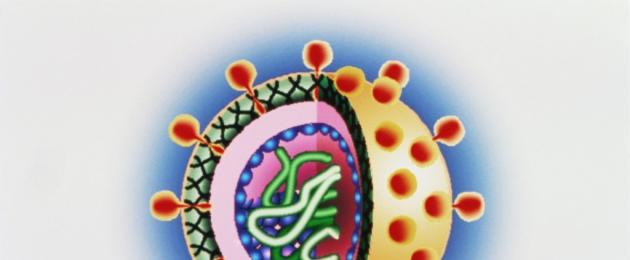
The morphology and structure of viruses is studied using an electron microscope, since their sizes are small and comparable to the thickness of the bacterial shell.
The shape of virions can be different: rod-shaped (tobacco mosaic virus), bullet-shaped (rabies virus), spherical (poliomyelitis viruses, HIV), sperm-shaped (many bacteriophages) (Fig. 8).
Rice. 8. Forms of virions:
1 smallpox virus; 2 herpes virus; 3 adenovirus; 4 papovavirus; 5 hepadnavirus; 6 paramyxovirus; 7 influenza virus; 8 coronavirus; 9 arenavirus; 10 retrovirus;
The sizes of viruses are determined using electron microscopy, ultrafiltration through filters with a known pore diameter, and ultracentrifugation. Some of the smallest viruses are polio and foot-and-mouth disease viruses (about 20 nm), circoviruses (16 nm), and the largest variola virus (about 350 nm). Viruses have a unique genome because they contain either DNA or RNA. Therefore, a distinction is made between DNA-containing and RNA-containing viruses. They are usually haploid, meaning they have one set of genes. The genome of viruses is represented by various types of nucleic acids: double-stranded, single-stranded, linear, circular, fragmented.
There are simple viruses (for example, polio virus) and complex viruses (for example, influenza viruses, measles viruses). In simple viruses, the nucleic acid is associated with a protein shell called a capsid (from the Latin capsa case). The capsid consists of repeating morphological subunits of capsomeres. Nucleic acid and capsid interact with each other to form a nucleocapsid. In complex viruses, the capsid is surrounded by an additional lipoprotein shell, a supercapsid (a derivative of the membrane structures of the host cell), which has “spikes”. The capsid and supercapsid protect virions from environmental influences, determine selective interaction (adsorption) with cells, and determine the antigenic and immunogenic properties of virions. The internal structures of viruses are called the core.
Virions are characterized by spiral, cubic and complex types of capsid symmetry. The helical type of symmetry is due to the helical structure of the nucleocapsid, the cubic formation of an isometric hollow body from the capsid containing the viral nucleic acid.
In addition to ordinary viruses, so-called non-canonical viruses are also known: prions, protein infectious particles that have the form of fibrils measuring 10-20 x 100-200 nm. Prions, apparently, are both inducers and products of an autonomous gene in humans or animals and cause encephalopathy in them under conditions of slow viral infection (Creutzfeldt-Jakob disease, kuru, etc.). Other unusual agents closely related to viruses are viroids, small circular, supercoiled RNA molecules that do not contain protein and cause diseases in plants.
3. Cultivation of viruses
Basic methods of cultivating viruses:
1) biological - infection of laboratory animals.
When an animal becomes infected with a virus, it becomes sick. If the disease is not
develops, then pathological changes can be detected
upon opening. Animals exhibit immunological
shifts. However, not all viruses can be cultivated
in the body of animals;
2) cultivation of viruses in developing chicken
embryos. Chicken embryos are grown in an incubator
7-10 days and then used for cultivation. In this
models, all types of tissue primordia are susceptible to infection.
But not all viruses can reproduce and develop in chickens.
ny embryos.
As a result of infection, the following may occur and appear:
1) death of the embryo;
2) developmental defects: appear on the surface of the membranes
formations - plaques, which are accumulations of
dead cells containing virions;
3) accumulation of viruses in allantoic fluid (detected
determined by titration);
4) reproduction in tissue culture (this is the main method of culture
virus titration).
The following types of tissue cultures are distinguished:
1) transplantable - tumor cell cultures; have
high mitotic activity;
2) primary trypsinized - subjected to primary
trypsin treatment; this treatment disrupts intercellular
connections, resulting in the separation of individual cells. Source
28
nickname are any organs and tissues, most often embryo-
nal (have high mitotic activity).
To maintain tissue culture cells, special
cial environments. These are liquid nutrient media of complex composition -
products containing amino acids, carbohydrates, growth factors, sources
protein nicknames, and indicators for assessing development
tissue culture cells.
The reproduction of viruses in tissue culture is judged by their cytology.
tactical action, which is of a different nature depending on
depending on the type of virus.
The main manifestations of the cytopathic effect of viruses:
1) the replication of the virus may be accompanied by the death of cells;
current or morphological changes in them;
2) some viruses cause cell fusion and the formation
formation of multinucleate syncytium;
3) cells can grow, but divide, resulting in the formation
there are giant cells;
4) inclusions appear in the cells (nuclear, cytoplasmic)
logical, mixed). Inclusions may appear pink
yellow color (eosinophilic inclusions) or blue (basic
philic inclusions);
5) if viruses that have
hemagglutinins, then during the process of reproduction the cell acquires
the ability to adsorb (hemadsorption) melts.
4. Features of antiviral immunity
Antiviral begins from the presentation stage
tions of viral T helper cells.
Strong antigen-presenting properties for virus-
In infections, dendritic cells have dendritic cells, and in simple germ-
dogs and retroviral infections - Langerhans cells.
aimed at neutralizing and removing from the organ-
nism of the virus, it and virus-infected cells. Anti-
bodies formed during viral infections act ineffectively
directly against the virus or cells infected by it. In this
communications identify two main forms of participation in development
antiviral immunity:
1) neutralization of the virus with antibodies; this interferes with the receptor
tion of the virus by the cell and its penetration inside. Opsonization
the virus promotes its phagocytosis;
29
2) immune lysis of virus-infected cells with the participation
eat . When antibodies act on antigens, expression
roved on the surface of the infected cell, to this
complement is added to the complex, followed by
activation, which determines the induction of complement-dependent
simultaneous cytotoxicity and death of the virus-infected
cells.
Insufficient concentration of antibodies can enhance repro-
virus duction. Sometimes antibodies can protect the virus from
effects of cell proteolytic enzymes, which, while maintaining
virus viability leads to increased replication.
Virus-neutralizing antibodies act directly
to the virus only in the case when it, having destroyed one cell, disintegrates
extends to another.
When viruses pass from cell to cell along the cytoplasmic
chemical bridges without contacting circulating antibodies,
then the main role in the formation of immunity is played by cellular
mechanisms associated primarily with the action of specific
cytotoxic, T-effectors and macrophages.
Cytotoxic directly contact
with the target cell, increasing its permeability and causing osmosis
tic swelling, membrane rupture and release of contents
into the environment.
The mechanism of the cytotoxic effect is associated with activation
membrane enzyme systems in the cell adhesion zone, forming
the formation of cytoplasmic bridges between cells and the action
the effect of lymphotoxin. Specific killer T cells appear
already 1-3 days after the body is infected with the virus, their ac-
activity peaks after a week and then slowly
goes down.
One of the factors of antiviral immunity is
interferon. It is formed in places where the virus multiplies and causes
causes specific inhibition of viral genome transcription
and suppression of viral mRNA translation, which prevents the accumulation
lysis of the virus in the target cell.
The durability of antiviral immunity is variable. When the
de infections (chicken pox, mumps, measles, rubella)
quite persistent, and recurrent diseases are extremely common
rarely. Less stable immunity develops during respiratory infections
chemical pathways (influenza) and intestinal tract.
- In contact with 0
- Google+ 0
- OK 0
- Facebook 0