Cells can move using specialized organelles, which include cilia and flagella. Cell cilia are always numerous (in protozoa their number is in the hundreds and thousands), and their length is 10-15 microns. There are most often 1-8 flagella, their length is 20-50 µm.
Structure and functions of movement organelles
The structure of cilia and flagella is similar in both plant and animal cells. Under an electron microscope, it was discovered that cilia and flagella are non-membrane organelles consisting of microtubules. Two of them are located in the center, and around them at the periphery lie another 9 pairs of microtubules. This entire structure is covered with a cytoplasmic membrane, which is a continuation of the cell membrane.
Flagella and cilia ensure not only the movement of cells in space, but also the movement of various substances on the surface of cells, as well as the entry of food particles into the cell. At the base of the cilia and flagella there are basal bodies, which also consist of microtubules.
It is believed that basal bodies are the center of formation of microtubules of flagella and cilia. Basal bodies, in turn, often originate from the cell center.
A large number of unicellular organisms and some multicellular cells do not have special organelles of movement and move with the help of pseudopodia (pseudopods), which is called amoeboid. It is based on the movement of molecules of special proteins called contractile proteins.
Features of the movement of protozoa
Single-celled organisms are also capable of moving (slipper ciliates, green euglena, common amoeba). To move through the water column, each individual is endowed with specific organelles. In protozoa, such organelles are cilia, flagella, and pseudopods.
Euglena green
Euglena green is a representative of the protozoa of the flagellate class. The body of euglena is spindle-shaped, elongated with a pointed end. The organelles of movement of Euglena greena are represented by a flagellum, which is located at the blunt end. Flagella are thin outgrowths of the body, the number of which varies from one to dozens.
The mechanism of movement using the flagellum differs in different species. Basically this is a rotation in the form of a cone, the top of which faces the body. The movement is most effective when the cone apex angle reaches 45°. The speed ranges from 10 to 40 revolutions per second. In addition to the rotational movement of the flagellum, its wave-like swaying is often observed.
This type of movement is typical for monoflagellate species. In polyflagellates, the flagella are often located in the same plane and do not form a cone of rotation.
The microscopic structure of flagella is quite complex. They are surrounded by a thin shell, which is a continuation of the outer layer of ectoplasm - the pellicle. The internal space of the flagellum is filled with cytoplasm and longitudinally arranged filaments - fibrils.
The peripherally located fibrils are responsible for movement, while the central fibrils perform a supporting function.
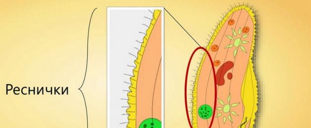
Ciliate slipper
The ciliate slipper moves due to the cilia, making wave-like movements with them. Points forward with the blunt end.
The cilia move in one plane and make a direct blow after full straightening, and a return blow in a curved position. The blows come sequentially one after another with a slight delay. While swimming, the ciliate performs rotational movements around the longitudinal axis.

The shoe moves at a speed of up to 2.5 mm/s. The direction changes due to the bends of the body. If there is an obstacle on the way, then after the collision the ciliate begins to move in the opposite direction.
All cilia of ciliates have a similar structure to the flagella of green euglena. The cilium at the base forms a basal grain, which plays an important role in the mechanism of movement of the body.
In some ciliates, the cilia are connected to each other and thus allow them to develop greater speed.
Ciliates are highly organized protozoa and they carry out their motor activity through contractions. The body shape of a protozoan can change and then return to its previous state. Rapid contractile movements are possible due to the presence of special fibers - myonemes.
Common amoeba
Amoeba is a fairly large protozoan (up to 0.5 mm). The body shape is polypodial, due to the presence of multiple pseudopodia - these are outgrowths with internal circulation of the cytoplasm.
In the common amoeba, the pseudopodia are also called pseudopods. Directing the pseudopods in different directions, the amoeba develops a speed of 0.2 mm/minute.
The organelles of protozoan movement do not include the cytoplasm, nucleus, vacuoles, ribosomes, lysosomes, ER, and Golgi apparatus.
Bacterial flagella determine the motility of the bacterial cell. Flagella are thin filaments originating from the cytoplasmic membrane and are longer than the cell itself. The thickness of the flagella is 12-20 nm, length 3-15 µm. They consist of 3 parts: a spiral filament, a hook and a basal body containing a rod with special disks (1 pair of disks in gram-positive bacteria and 2 pairs of disks in gram-negative bacteria). Flagella are attached to the cytoplasmic membrane and cell wall by discs. This creates the effect of an electric motor with a motor rod that rotates the flagellum. Flagella consist of a protein - flagellin (from flagellum - flagellum); is an H antigen. Flagellin subunits are twisted in a spiral. The number of flagella in bacteria of various species varies from one (monotrich) in Vibrio cholerae to tens and hundreds of flagella extending along the perimeter of the bacterium (peritrich) in Escherichia coli, Proteus, etc. Lophotrichs have a bundle of flagella at one end of the cell. Amphitrichy has one flagellum or a bundle of flagella at opposite ends of the cell.
Pili (fimbriae, villi) are thread-like formations, thinner and shorter (3-10 nm x 0.3-10 µm) than flagella. Pili extend from the cell surface and consist of the protein pilin, which has antigenic activity. There are pili responsible for adhesion, that is, for attaching bacteria to the affected cell, as well as pili responsible for nutrition, water-salt metabolism and sexual (F-pili), or conjugation pili. Pili are numerous - several hundred per cell. However, there are usually 1-3 sex pili per cell: they are formed by so-called “male” donor cells containing transmissible plasmids (F-, R-, Col-plasmids). A distinctive feature of the sex pili is the interaction with special “male” spherical bacteriophages, which are intensively adsorbed on the sex pili.
Spores are a peculiar form of resting firmicute bacteria, i.e. bacteria with a gram-positive type of cell wall structure. Spores are formed under unfavorable conditions for the existence of bacteria (drying, nutrient deficiency, etc.. One spore (endospore) is formed inside the bacterial cell. The formation of spores contributes to the preservation of the species and is not a method of reproduction, like fungi. Spore-forming bacteria of the genus Bacillus have spores, not exceeding the diameter of the cell. Bacteria in which the size of the spore exceeds the diameter of the cell are called clostridia, for example, bacteria of the genus Clostridium (lat. Clostridium - spindle). in blue.
The shape of the spores can be oval, spherical; location in the cell is terminal, i.e. at the end of the stick (in the causative agent of tetanus), subterminal - closer to the end of the stick (in the causative agents of botulinum, gas gangrene) and central (in the anthrax bacillus). The spore persists for a long time due to the presence of a multilayer shell, calcium dipicolinate, low water content and sluggish metabolic processes. Under favorable conditions, spores germinate, going through three successive stages: activation, initiation, germination.
8. Basic forms of bacteria
Globular bacteria (cocci) They are usually spherical in shape, but can be slightly oval or bean-shaped. Cocci can be located singly (micrococci); in pairs (diplococci); in the form of chains (streptococci) or grape bunches (staphylococci), in a package (sarcins). Streptococci can cause tonsillitis and erysipelas, while staphylococci can cause various inflammatory and purulent processes.
Rod-shaped bacteria the most common. The rods can be single, connected in pairs (diplobacteria) or in chains (streptobacteria). The rod-shaped bacteria include Escherichia coli, the causative agents of salmonellosis, dysentery, typhoid fever, tuberculosis, etc. Some rod-shaped bacteria have the ability to form disputes. Spore-forming rods are called bacilli. Spindle-shaped bacilli are called clostridia.
Sporulation is a complex process. Spores are significantly different from an ordinary bacterial cell. They have a dense shell and a very small amount of water, they do not require nutrients, and reproduction completely stops. Spores are able to withstand drying, high and low temperatures for a long time and can remain in a viable state for tens and hundreds of years (spores of anthrax, botulism, tetanus, etc.). Once in a favorable environment, the spores germinate, that is, they turn into the usual vegetative propagating form.
Twisted bacteria can be in the form of a comma - vibrios, with several curls - spirilla, in the form of a thin twisted stick - spirochetes. Vibrios include the causative agent of cholera, and the causative agent of syphilis is a spirochete.
9. Features of the morphology of rickettsia and chlamydia
Rickettsia are small gram-negative microorganisms characterized by pronounced polymorphism - they form coccoid, rod-shaped and filamentous forms (Fig. 22). The size of rickettsia varies from 0.5 to 3-4 microns, the length of filamentous forms reaches 10-40 microns. They do not form spores or capsules and are stained red according to Zdrodovsky.
Chlamydia are spherical, ovoid or rod-shaped. Their sizes range from 0.2-1.5 microns. The morphology and size of chlamydia depend on the stage of their intracellular development cycle, which is characterized by the transformation of a small spherical elementary formation into a large initial body with binary fission. Before dividing, chlamydia particles are enveloped in a formation resembling a bacterial capsule. Chlamydia are stained* according to Romanovsky-Giemsa, are gram-negative, and are clearly visible in intravital specimens with phase-contrast microscopy.
10. Structure and biology of mycoplasmas.
The class Mollicutes includes only one order, Mycoplasmatales. Representatives of this order - mycoplasma -
They differ from bacteria in that they lack a cell wall. Instead, they contain a three-layer lipoprotein cytoplasmic membrane. The sizes of mycoplasmas range from 125-250 microns. They have the shape of round, oval or filamentous formations, gram-negative
Mycoplasmas reproduce by binary fission, like most bacteria, especially after the formation of small coccoid formations (elementary bodies, EB) in filamentous structures.
Mycoplasmas are capable of budding and segmentation. The minimum reproducing unit is considered to be the ET (0.7-0.2 µm). The main component of the cell membrane is cholesterol. Mycoplasmas are not capable of producing cholesterol and utilize it from tissues or nutrient media supplemented with their addition. The Gram stain is negative, but the best results are obtained by Romanowsky-Giemsa stain. Mycoplasmas are demanding regarding cultivation conditions: native serum, cholesterol, nucleic acids, carbohydrates, vitamins and various salts must be added to the nutrient media. On dense media they form characteristic small translucent colonies with a raised granular center, giving them a “fried egg” appearance. On media containing blood, some types of mycoplasmas produce a- and beta-hemolysis. In semi-liquid media, mycoplasmas grow along the injection line, forming dispersed, crumbly colonies. In liquid media they lead to slight turbidity or opalescence; some strains are capable of forming a thin oily film. In humans, representatives of the genera Mycoplasma, Ureaplasma and Acholeplasma are isolated, including pathogenic and saprophytic species
All bacteria are divided into movable and immobile. The organs of movement in bacteria are flagella. They consist of the protein flagellin, which in its structure belongs to the contractile proteins of the myosin type.
Base of flagellum is a basal body, consisting of a system of disks (blepharoplast: 1 disk - the outer side of the cell wall, 2 disk - the inner side of the cell wall, 3 disk - the cytoplasmic membrane), “built in” into the cytoplasmic membrane and the cell wall. The length of the flagellum is greater than the length of the body of the microbe itself.
According to the number of flagella and their location, motile microorganisms are divided into:
1. Monotrichs, which have one flagellum at the end of the body (the most mobile). For example, Vibrio cholerae.
2. Lophotrichs, which have a bundle of flagella at one of the cell poles. For example, Burkholderia (Pseudomonas) pseudomalei is the causative agent of melioidosis.
3. Amphitrichy, which has a flagellum at both poles of the cell. For example, Spirillum volutans.
4. Peritrichs, which have flagella along the entire perimeter of the cell. For example, Escherichia coli, Salmonella typhi.
Identification of flagella. The flagella are very thin, so they can only be detected with special processing. In particular, first, with the help of a mordant, swelling and an increase in their size are achieved, and then the preparation is colored, due to which they become visible under light microscopy. Flagella can be identified by Morozov, Leffler staining, as well as electron microscopy. Flagella can also be detected by the active motility of bacteria.
The movement of microbes is observed in “crushed” and “hanging” drop preparations from living cultures. These preparations are microscoped with a dry or immersion objective in a dark field or in phase contrast. In addition, motility can be determined by the growth pattern of bacteria in semi-solid agar.
They drank from bacteria.
Pili (pili), synonyms: villi, fimbriae, are thin hollow filaments of a protein nature that cover the surface of bacterial cells. Unlike flagella, they do not perform a motor function.
Pili extend from the surface of the cell and are made of protein pilina.
According to their functional purpose they are divided into 2 types.
1) Most bacteria have pili of the first type, which is why they are called “common pili”. They cause the attachment or adhesion of bacteria to certain cells of the host body. Adhesion is the initial stage of any infectious process.
2) Pili of the second type (synonyms: conjugative, or sexual - sex pili) are found only in donor bacteria that have a special plasmid. Their number is small - 1-4 per cell.
Sex saws perform the following functions:
1. Participate in the transfer of genetic material from one cell to another during the conjugation of bacteria.
2. Specific bacterial viruses – bacteriophages – are adsorbed on them
Bacterial spores, conditions of formation, location, mechanism and stages of Aujeszky staining.
Controversy- a peculiar form of resting bacteria with a gram-positive type of cell wall structure.
Sporulation- this is a way of preserving a species (genophore) in the external environment under unfavorable conditions, and not a method of reproduction.
Spores are formed under unfavorable conditions for the existence of bacteria (drying, nutrient deficiency, etc.). A single spore (endospore) is formed inside a bacterial cell.
Stages of sporulation
1. Preparatory. In the cytoplasm of bacteria, a compacted area is formed that does not have free water, called the “sporogenic zone,” which contains the nucleoid.
2. Prespore stage (prospores). A shell of a double cytoplasmic membrane is formed around the sporogenic zone.
3. Formation of a cortex consisting of peptidoglycan and an outer membrane with a high content of calcium salts and lipids.
4. Maturation stage. A spore shell is formed on the outside of the outer membrane, after which the vegetative part of the cell lyses, releasing the spore.
There are a large number of microbes with flagella. The flagella of bacteria are their characteristic features, and according to this principle they are combined into taxonomic units. Thanks to the processes, these organisms are able to contract the cell and thus move.
These structural elements of the cell determine its mobility. Most often these are thin filaments that originate from the cytoplasmic membrane. Some types of microbes have a significantly larger flagellum than the host cell itself.
The processes are capable of pushing the cell through a liquid medium. The structure of the flagellum is such that it can quickly move the cell body, and at the same time it will cover relatively long distances. These movements are performed according to the principle of a propeller. To move, microbes use one or more processes.
In some microbes, processes can be an additional factor of pathogenicity (pathogenicity). This can be explained by the fact that it helps the pathogenic microorganism approach the healthy cell.
What are flagella made of?
These parts of the microorganism are spirally twisted threads. They have different thicknesses and lengths, as well as coil amplitudes. Some bacteria with flagella have several varieties of these organs.
These cell elements consist of a special protein – flagellin. It has a relatively small molecular weight. This allows the subunits of the molecules to be arranged in a spiral and thus form the structure of a process of a certain length.

In addition to the filament, the tourniquet has a hook near the surface of the cell, as well as a basal body. With the help of such a body, it is securely attached to the cell.
What are villi
The villi are otherwise called pili. They are present in different organisms. The arrangement of these structural elements of the bacterial cell is different. Typically these are cylinders of protein nature, having a length of up to 1.5 micrometers and a diameter of up to 1 micrometer. One microorganism can contain several types of pili.
The functions of these formations have not yet been fully determined. It is known that certain types of microbes have villi. The most obvious role played by pili is attachment to the substrate and movement in the environment.
The most data has been collected on E. coli, which have pili. However, there are a huge number of microscopic organisms in which the structure of the villi has not yet been fully determined. In any case, bacterial pili promote efficient cell movement.
What are the differences between flagellated microorganisms?
Depending on the number and method of arrangement, all microscopic organisms are divided into the following types:
- Monotrichs. These are bacteria with one flagellum.
- Lophotrichs. These cells have a bundle of processes at the end.
- Peritrichous. Such microbes have many processes over the entire surface.
- Amphitrichy. These microorganisms have a bilateral, or bipolar, arrangement of flagella.
Flagella of prokaryotes
In prokaryotic bacteria, such elements consist of only one region of flagellin subunits. One- or two-sided arrangement of such elements is possible. To a large extent, such parts of the cell can be determined by differences in the life cycle.
Some prokaryotic bacteria may have pili. The number of these structural elements allows the bacterium to move or attach to the substrate.
Most prokaryotes have excellent adaptations to move in a liquid environment and thereby increase survival under unfavorable environmental factors.
Eukaryotic flagella

Flagella in eukaryotic microorganisms are much thicker and have a complex structure. Unlike prokaryotic microorganisms, these bacteria with flagella can rotate independently. The pili in such organisms give them the ability to additionally attach to the substrate, as well as perform complex movements.
In some microorganisms, flagella have a more complex structure - in the form of microtubules. Such a tube has tightly packed strands of protein molecules. They excel at moving in a variety of environments. Microtubules apparently arose at the later stages of the evolution of microorganisms.
How to identify flagella
Conventionally, flagella can be determined by direct and indirect methods.
Observing bacteria through a microscope is a direct detection of these elements. To make them more noticeable, special staining methods are used. The flagella are even better visible under an electron microscope.
Indirectly, bacteria are determined by the fact of cell motility. This is best detected using the “crushed drop” preparation, when the slide is covered with a coverslip. Often, in order to make the processes more visible, the field of view is artificially darkened.
The study of flagellated bacteria and their functions allows microbiologists to find ways to combat pathogens, as well as fields for their application.

I work as a veterinary doctor. I am interested in ballroom dancing, sports and yoga. I prioritize personal development and mastering spiritual practices. Favorite topics: veterinary medicine, biology, construction, repairs, travel. Taboos: law, politics, IT technologies and computer games.
Table of contents of the topic "Anatomy of a bacterial cell. Physiology of bacteria.":1. Anatomy of a bacterial cell. Surface structures of bacteria. Bacteria capsule. Organization of capsules. Staining of bacterial capsules. Composition of capsules. Antigenic properties of capsules.
3. Microvilli of bacteria. Fimbriae of bacteria. F-pili (sex-pili) bacteria. Cell membrane of bacteria. Glycocalyx.
4. Cell wall of bacteria. Functions of the cell wall. The structure of the bacterial cell wall. Peptidoglycan. Murein sac. Structure of peptidoglycan (murein)
5. Gram-negative bacteria. Cell wall of gram-negative bacteria. The structure of the cell wall of gram-negative bacteria.
6. Gram-positive bacteria. Cell wall of gram-positive bacteria. The structure of the cell wall of gram-positive bacteria. Bacterial autolysins. Spheroplasts. Protoplasts.
7. Cytoplasmic membrane (CPM) of bacteria. Composition of the bacterial cytoplasmic membrane. Transport systems. Mesosomes. Periplasmic space.
8. Cytoplasm of bacteria. Bacterial genome. Bacterial ribosomes. Spare bacterial granules.
9. Physiology of bacteria. Nutrition of bacteria. Type of nutrition of bacteria. Holozoans. Holophytes. Water. The importance of water for bacteria.
10. Compounds assimilated by the bacterial cell. Pathways for substances to enter the bacterial cell. Passive transfer. Diffusion.
By nature of movement motile bacteria divided into floating And sliding(crawling). Organ of movement of floating bacteria - flagella; The mobility of sliding bacteria is ensured by wave-like contractions of the body.
Location of flagella- a characteristic feature that has taxonomic significance. Variants of the location of flagella are shown in Fig. 4-1. In some bacteria, flagella are located over the entire surface of the cell wall (for example, in bacteria of the genus Proteus); such bacteria are known as peritrichous[from Greek peri-, around, + trichos, hair]. Some bacteria are equipped with only one thick flagellum (for example, representatives of the genus Vibrio), they are known as monotrichs. Polytrichs- bacteria that have a single flagellum formed by a bundle of 2-50 flagella. Polar flagella are attached to one or both ends of the bacterium. The flagella have a monopolar-polytrichial arrangement lophotrichs[from Greek lophos, tuft, + trichos, hair], these include, for example, representatives of the genus Pseudomonas. Bipolar-polytrichial flagellation has amphitrichs[from Greek amphi-, bilateral, + trichos, hair] (for example, bacteria of the genus Spirillum).
Rice. 4-1. Variants of the location of flagella (top) and bacterial movements (bottom).Flagellum- a spirally curved hollow filament formed by flagellin subunits. In different bacteria, the thickness of the flagella varies from 12 to 18 nm, which is no more than 1/10 of the diameter of the flagella of algae and protozoa. Flagella are also distinguished by the length and diameter of the coil. The place of attachment of the flagellum to the bacterial cell has a complex structure and consists of a basal structure and the so-called “hook” (Fig. 4-2). In gram-positive bacteria, the basal structure includes one pair, and in gram-negative bacteria, two pairs of rings. The rings play the role of “drive disk” and “bearing”. The entire structure performs the function of a chemomechanical converter (flagellin motor). In spirochetes, a special organelle is responsible for movement - an axial filament, consisting of two rows of bacterial flagella located longitudinally inside the cell.
Bacterial flagella perform translational and rotational movements, pushing bacteria through the medium like a ship's propeller. They can also change the direction of rotation and pull the cage like a propeller. The speed of reverse movement is four times less than the speed of forward movement. Some peritrichs can move on the surface of agar, that is, floating bacteria are capable of moving on the surface of solid media. In particular, Proteus vulgaris spreads over the surface of the agar, forming a thin coating (reminiscent of that when exhaling on cold glass), and non-motile strains of Proteus lack this ability. This phenomenon is called " swarming phenomenon", and observation of it formed the basis of some concepts of bacterial serodiagnosis. Thus, flagellated Ags are called N-Ars [from German. Hauch, exhalation, raid], and Ag of the cell surface - O-Ar [from German. FPE Hauch, without flying time1.
 Rice. 4-2. Scheme of the structure of a bacterial flagellum. BS - basal structure, VM - outer membrane, CPM - cytoplasmic membrane, R - rotor, O - axis, KO - flagellar motor ring, KR - hook, C - connector cylinders, N - flagellar filament, W - cap.
The ability of bacteria to target movement genetically determined. For example, in Escherichia coli, 3% of the genome (approximately 50 genes) is involved in the regulation of this process. These genes encode proteins that form the locomotor apparatus, as well as proteins and enzymes involved in signal transduction. For the flagellar apparatus characterized by periodic variability. In many ways, this process is adaptive in nature and is most pronounced in pathogenic microorganisms. In particular, some bacteria have developed a system of variability in the antigenic characteristics of flagella, allowing them to temporarily avoid the targeted effects of protective immune mechanisms.
Laboratory diagnostics of bacterial motility
Motility of bacteria determined by microscopy of preparations in “ crushed" or " hanging» drop. The ability to move can also be determined after introducing a bacterial culture by injection into a column of semi-liquid agar (mobile species grow throughout the entire thickness of the medium, immobile species by injection) or by sowing bacteria into the water condensate of a slanted agar column (mobile species swim from the condensate to the surface of the medium and colonize it ), or determine the ability of bacteria to produce " swarming phenomenon».
- In contact with 0
- Google+ 0
- OK 0
- Facebook 0