The simplest animals are single-celled organisms, characteristics, nutrition, presence in water and in the human body
General characteristics
Or unicellular organisms, as their name suggests, are made up of a single cell. The phylum Protozoa includes more than 28,000 species. The structure of protozoa can be compared with the structure of cells of multicellular organisms. Both of them are based on the nucleus and cytoplasm with various organelles (organelles) and inclusions. However, we must not forget that any cell of a multicellular organism is part of any tissue or organ where it performs its specific functions. All cells of a multicellular organism are specialized and are not capable of independent existence. In contrast, the simplest animals combine the functions of a cell and an independent organism. (Physiologically, the Protozoa cell is similar not to individual cells of multicellular animals, but to a whole multicellular organism.
The simplest all functions inherent in any living organisms are characteristic: nutrition, metabolism, excretion, perception of external stimuli and reaction to them, movement, growth, reproduction and death.
Protozoa Cell structure
The nucleus and cytoplasm, as indicated, are the main structural and functional components of any cell, including unicellular animals. The body of the latter contains organelles, skeletal and contractile elements and various inclusions. It is always covered with a cell membrane, more or less thin, but clearly visible in an electron microscope. The cytoplasm of protozoa is liquid, but its viscosity varies among different species and varies depending on the condition of the animal and the environment (its temperature and chemical composition). In most species the cytoplasm is transparent or milky white, but in some it is colored blue or greenish (Stentor, Fabrea saliva). The chemical composition of the nucleus and cytoplasm of protozoa has not been fully studied, mainly due to the small size of these animals. It is known that the basis of the cytoplasm and nucleus, as in all animals, is made up of proteins. Nucleic acids are closely related to proteins; they form nucleoproteins, the role of which in the life of all organisms is extremely large. DNA (deoxyribonucleic acid) is part of the chromosomes of the protozoan nucleus and ensures the transmission of hereditary information from generation to generation. RNA (ribonucleic acid) is found in protozoa both in the nucleus and in the cytoplasm. It implements the hereditary properties of single-celled organisms encoded in DNA, as it plays a leading role in the synthesis of proteins.
Very important chemical components of the cytoplasm - fat-like substances lipids - take part in metabolism. Some of them contain phosphorus (phosphatides), many are associated with proteins and form lipoprotein complexes. The cytoplasm also contains reserve nutrients in the form of inclusions - droplets or granules. These are carbohydrates (glycogen, paramyl), fats and lipids. They serve as the energy reserve of the protozoan body.
In addition to organic substances, the cytoplasm contains a large amount of water and mineral salts (cations: K+, Ca2+, Mg2+, Na+, Fe3+ and anions: Cl~, P043“, N03“). In the cytoplasm of protozoa, many enzymes involved in metabolism are found: proteases, which ensure the breakdown of proteins; carbohydrases that break down polysaccharides; lipases that promote fat digestion; a large number of enzymes that regulate gas exchange, namely alkaline and acid phosphatases, oxidases, peroxidases and cytochrome oxidases.
Previous ideas about the fibrillar, granular or foamy-cellular structure of the cytoplasm of protozoa were based on studies of fixed and stained preparations. New methods for studying protozoa (in a dark field, in polarized light, using intravital staining and electron microscopy) have made it possible to establish that the cytoplasm of protozoa is a complex dynamic system of hydrophilic colloids (mainly protein complexes), which has a liquid or semi-liquid consistency. During ultramicroscopic examination in a dark field, the cytoplasm of protozoa appears optically empty, only the cell organelles and its inclusions are visible.
The colloidal state of cytoplasmic proteins ensures the variability of its structure. In the cytoplasm, changes in the aggregate state of proteins constantly occur: they pass from a liquid state (sol) to a more solid, gelatinous state (gel). These processes are associated with the release of a denser layer of ectoplasm, the formation of a shell - pellicles, and the amoeboid movement of many protozoa.
The nuclei of protozoa, like the nuclei of multicellular cells, consist of chromatin material, nuclear juice, and contain nucleoli and a nuclear envelope. Most protozoa contain only one nucleus, but there are also multinucleate forms. In this case, the nuclei can be the same (multinucleate amoebas from the genus Pelomyxa, multinucleate flagellates Polymastigida, Opalinida) or differ in shape and function. In the latter case, they talk about nuclear differentiation, or nuclear dualism. Thus, the entire class of ciliates and some foraminifera are characterized by nuclear dualism. i.e. nuclei unequal in shape and function.
These types of protozoa, like other organisms, obey the law of constancy of the number of chromosomes. Their number can be single, or haploid (most flagellates and sporozoans), or double, or diploid (ciliates, opalines and, apparently, sarcodae). The number of chromosomes in different species of protozoa varies widely: from 2-4 to 100-125 (in the haploid set). In addition, nuclei with a multiple increase in the number of sets of chromosomes are observed. They are called polyploid. It was found that large nuclei, or macronuclei, of ciliates and the nuclei of some radiolarians are polyploid. It is very likely that the nucleus of Amoeba proteus is also polyploid; the number of chromosomes in this species reaches 500.
Reproduction Nuclear division
The main type of nuclear division in both protozoa and multicellular organisms is mitosis, or karyokinesis. During mitosis, correct, uniform distribution of chromosomal material occurs between the nuclei of dividing cells. This is ensured by the longitudinal splitting of each chromosome into two daughter chromosomes in the metaphase of mitosis, with both daughter chromosomes going to different poles of the dividing cell.
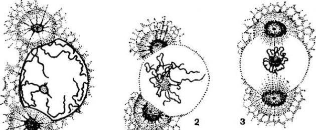
 Mitotic division of the gregarine nucleus of Monocystis magna:
 1, 2 - prophase; 3 - transition to metaphase; 4, 5 - metaphase; 6 - early anaphase; 7, 8 - late
anaphase; 9, 10 - telophase.
When the nucleus of Monocystis magna gregarina divides, all the mitotic figures characteristic of multicellular organisms can be observed. In prophase, thread-like chromosomes are visible in the nucleus, some of them are associated with the nucleolus (Fig. 1, 1, 2). In the cytoplasm, two centrosomes can be distinguished, in the center of which there are centrioles with star rays diverging radially. Centrosomes approach the nucleus, adjoin its shell and move to the opposite poles of the nucleus. The nuclear envelope dissolves and an achromatin spindle is formed (Fig. 1, 2-4). Spiralization of chromosomes occurs, as a result of which they are greatly shortened and collected in the center of the nucleus, the nucleolus dissolves. In metaphase, chromosomes move to the equatorial plane. Each chromosome consists of two chromatids lying parallel to each other and held together by one centromere. The star figure around each centrosome disappears, and the centrioles are divided in half (Fig. 1, 4, 5). In anaphase, the centromeres of each chromosome divide in half and their chromatids begin to diverge towards the spindle poles. It is characteristic of protozoa that the pulling spindle filaments attached to the centromeres are distinguishable only in some species. The entire spindle is stretched, and its threads, running continuously from pole to pole, lengthen. The separation of chromatids that have turned into chromosomes is ensured by two mechanisms: their pulling apart under the action of contraction of the pulling spindle threads and the stretching of continuous spindle threads. The latter leads to the removal of the cell poles from each other (Fig. 1, 6, 7). In telophase, the process occurs in the reverse order: at each pole, a group of chromosomes is clothed with a nuclear envelope. The chromosomes despiral and become thinner, and nucleoli are formed again. The spindle disappears, and around the divided centrioles two independent centrosomes with star rays are formed. Each daughter cell has two centrosomes - the future centers of the next mitotic division (Fig. 1, 9, 10). However, in some protozoa the cytoplasm also divides. , including in Monocystis, a series of successive nuclear divisions occur, as a result of which temporarily multinuclear stages appear in the life cycle. Later, a section of cytoplasm is isolated around each nucleus and many small cells are formed simultaneously.
There are various deviations from the process of mitosis described above: the nuclear envelope can be preserved throughout the entire mitotic division, the achromatin spindle can form under the nuclear envelope, and in some forms centrioles are not formed. The most significant deviations are in some euglenidae: they lack a typical metaphase, and the spindle passes outside the nucleus. In metaphase, chromosomes, consisting of two chromatids, are located along the axis of the nucleus, the equatorial plate is not formed, the nuclear membrane and nucleolus are preserved, the latter is divided in half and passes into the daughter nuclei. There are no fundamental differences between the behavior of chromosomes in mitosis in protozoa and multicellular organisms.
Before the use of new research methods, the nuclear division of many protozoa was described as amitosis, or direct division. True amitosis is now understood as the division of nuclei without proper separation of chromatids (chromosomes) into daughter nuclei. As a result, nuclei with incomplete sets of chromosomes are formed. They are not capable of further normal mitotic divisions. It is difficult to expect such nuclear divisions in the simplest organisms normally. Amitosis is observed optionally as a more or less pathological process.
The body of protozoa is quite complex. Within one cell, differentiation of its individual parts occurs, which perform different functions. Thus, by analogy with the organs of multicellular animals, these parts of protozoa were called organelles or organelles. There are organelles of movement, nutrition, perception of light and other stimuli, excretory organelles, etc.
Movement
The organelles of movement in Protozoa are pseudopodia, or pseudopods, flagella and cilia. Pseudopodia are formed for the most part at the moment of movement and can disappear as soon as the protozoan stops moving. Pseudopodia are temporary plasmatic outgrowths of the body of protozoa that do not have a permanent shape. Their shell is represented by a very thin (70-100 A) and elastic cell membrane. Pseudopodia are characteristic of sarcodae, some flagellates and sporozoans.
Flagella and cilia are permanent outgrowths of the outer layer of the cytoplasm, capable of rhythmic movements. The ultrafine structure of these organelles was studied using an electron microscope. It was found that they are constructed in much the same way. The free part of the flagellum or cilium extends from the surface of the cell.
The internal part is immersed in ectoplasm and is called the basal body or blepharoplast. On ultrathin sections of a flagellum or cilium, 11 longitudinal fibrils can be distinguished, 2 of which are located in the center, and 9 along the periphery (Fig. 2). The central fibrils in some species have helical striations. Each peripheral fibril consists of two connected tubes, or subfbrils. Peripheral fibrils pass into the basal body, but central fibrils do not reach it. The flagellum membrane passes into the membrane of the protozoan body.
Despite the similarity in structure of cilia and flagella, the nature of their movement is different. If flagella make complex screw movements, then the work of cilia can most easily be compared with the movement of oars.
In addition to the basal body, the cytoplasm of some protozoa contains a parabasal body. The basal body is the basis of the entire musculoskeletal system; in addition, it regulates the process of mitotic division of the protozoan. The parabasal body plays a role in the metabolism of the protozoan; at times it disappears and then may appear again.
Sense organs
Protozoa have the ability to determine light intensity (illuminance) using a photosensitive organelle - the ocellus. A study of the ultrathin structure of the eye of the sea flagellate Chromulina psammobia showed that it includes a modified flagellum immersed in the cytoplasm.
In connection with the different types of nutrition, which will be discussed in detail later, protozoa have a very wide variety of digestive organelles: from simple digestive vacuoles or vesicles to such specialized formations as the cellular mouth, oral funnel, pharynx, powder.
Excretory system
Most protozoa are characterized by the ability to withstand unfavorable environmental conditions (drying out of temporary reservoirs, heat, cold, etc.) in the form of cysts. In preparation for encystment, the protozoan releases a significant amount of water, which leads to an increase in the density of the cytoplasm. The remains of food particles are thrown out, the cilia and flagella disappear, and the pseudopodia are retracted. The overall metabolism decreases, a protective shell is formed, often consisting of two layers. The formation of cysts in many forms is preceded by the accumulation of reserve nutrients in the cytoplasm.
Protozoa do not lose viability in cysts for a very long time. In experiments, these periods exceeded 5 years for the genus Oicomonas (Protomonadida), 8 years for Haematococcus pluvialis, and for Peridinium cinctum the maximum survival period of cysts exceeded 16 years.
In the form of cysts, protozoa are transported by wind over considerable distances, which explains the homogeneity of the protozoan fauna throughout the globe. Thus, cysts not only have a protective function, but also serve as the main means of dispersal of protozoa.
Covers of the body.
Body shape, symmetry.
The body shape of protozoa and its color are extremely diverse and determined by specific living conditions. Functionally, the anterior end of the flagellate is where the flagellum is attached.
All protozoa, regardless of their type of organization, are protected from the effects of the external environment by cell membranes of various structures. The main structural unit of all types of integument in protozoa is the cytoplasmic membrane. On the inner side of the plasmalemma there are usually submembrane microfilaments or microtubules.
The appearance of flagella as a locomotor apparatus led to the appearance of relatively another type of integument in flagellates - dense pellicles. The pellicle is formed due to the compaction of the peripheral layer of the cytoplasm and the presence of supporting fibrils in it. It is strengthened by outgrowths of the radicular system.
The next stage in the complication of the integument is the external skeleton, formed by protein, cellulose and even chitinous plates, calcareous, silica structures, as well as glycoprotein gelatinous secretions in some flagellates.
In some protozoa, the integument of various types is complicated by the appearance of a more or less complex sculpture, that is, a system of more or less regularly located depressions and projections that form something like stiffening ribs (Opalinidomorpha), “reinforced” with microtubules. Such covers are called folded or comb tubulemma.
Characteristic of ciliates cortex. The cortex includes: a pellicle (formed by a membrane and a system of alveoli), under the pellicle there is a protein layer - epiplasm and a complex of kinetosomes.
TO general cellular structures include: cytoplasm, nucleus, mitochondria, endoplasmic reticulum, ribosomes, lysosomes, Golgi apparatus, centriole.
One core or several of them. Depending on the number of nuclei, protozoa are divided into monoenergetic and polyenergetic. Ciliates are characterized by nuclear dualism: the functions of the nuclei (micronucleus and macronucleus) differ.
Special organelles cells are: contractile and digestive vacuoles, microfilaments (participate in the processes of contraction and cell division, form fibrils), microtubules (the main function is the formation of the cytoskeleton, take part in cell division, in the formation of the oral apparatus, hold organelles in a certain position), extrusomes ( varied in shape, in response to irritation they throw out the contents), powder, stigma, flagella and cilia.
Inclusions are: droplets of fat, protein crystals, symbiotic organisms.
In the middle part of the body sporozoite there is a round hole, micropyle. It is known that after the sporozoite penetrates the cell of a vertebrate host, it immediately becomes rounded, which is difficult to explain, given the density of its pellicle. Garnham and co-workers (1963) suggested that the sporozoite cytoplasm with the nucleus at this moment emerges through the micropyle, from the shell.
Installed similarity in structure sporozoites and merozoites; in blood trophozoites, a micropyle-like formation has been described, possibly serving as a cytostome (Aikawa, 1966).
Digestion and absorption of food in protozoa it occurs in digestive vacuoles - vesicles located in the inner layer of the cytoplasm. The food for protozoa can be both formed particles (including various living organisms) and substances dissolved in the environment. The absorption of formed particles is carried out through phagocytosis. Their capture in sarcodes occurs at any point on the surface.
However, a significant part protozoa For this process, any one part of the body is specialized: a depression is formed - the cell mouth of the cytostome. The structure of the centostome of ciliates is especially complex, in which it can be surrounded by special organelles - elongated cilia and membranella.
Absorption dissolved nutrients carried out through the capture of environmental droplets by the surface of the cytoplasm. The resulting vesicles pass through the cell membrane, detach from it and move into the cytoplasm. This process is called pinocytosis.
Every living organism is made up of cells, many of which are capable of movement. In this article we will talk about movement organelles, their structure and functions.
Organelles of movement of unicellular organisms
In modern biology, cells are divided into prokaryotes and eukaryotes. The first include representatives of the simplest organisms that contain one strand of DNA and do not have a nucleus (blue-green algae, viruses).
Eukaryotes have a nucleus and consist of a variety of organelles, one of which is the organelles of movement.
Organelles of movement of unicellular organisms include cilia, flagella, thread-like formations - myofibrils, pseudopods. With their help, the cell can move freely.

Rice. 1. Varieties of movement organelles.
Movement organelles are also found in multicellular organisms. For example, in humans, the bronchial epithelium is covered with many cilia, which move strictly in the same order. In this case, a so-called “wave” is formed that can protect the respiratory tract from dust and foreign particles. Spermatozoa (specialized cells of the male body that serve for reproduction) also have flagella.
TOP 4 articleswho are reading along with this
The motor function can also be achieved due to the contraction of microfibers (myonemes), which are located in the cytoplasm under the integument.
Structure and functions of movement organelles
Movement organelles are membrane outgrowths that reach 0.25 µm in diameter. In terms of their structure, flagella are much longer than cilia.
The length of the sperm flagellum in some mammals can reach 100 microns, while the size of the cilia is up to 15 microns.
Despite such differences, the internal structure of these organelles is absolutely the same. They are formed from microtubules, which are similar in structure to the centrioles of the cell center.
Motor movements are formed due to the sliding of microtubules among themselves, as a result of which they bend. At the base of these organelles there is a basal body that attaches them to the cell cytoplasm. To ensure the functioning of movement organelles, the cell consumes ATP energy.

Rice. 2. Structure of the flagellum.
Some cells (amoebas, leukocytes) move due to pseudopodia, in other words, pseudopods. However, unlike flagella and cilia, pseudopodia are temporary structures. They can disappear and appear in different places in the cytoplasm. Their functions include locomotion and the capture of food and other particles.
Flagella consist of a filament, a hook and a basal body. According to the number and location of these organelles on the surface of bacteria they are divided into:
- Monotrichs(one flagellum);
- Amphitrichy(one flagellum at different poles);
- Lophotrichs(a bunch of formations on one or both poles);
- Peritrichous(many flagella located over the entire surface of the cell).

Rice. 3. Varieties of flagellates.
Among the functions performed by movement organelles are:
- providing movement to a single-celled organism;
- the ability of muscles to contract;
- protective reaction of the respiratory tract from foreign particles;
- fluid advancement.
Flagellates play a large role in the cycle of substances in the environment; many of them are good indicators of pollution of water bodies.
What have we learned?
One of the constituent elements of the cell are organelles of movement. These include flagella and cilia, which are formed with the help of microtubules. Their functions include providing movement to a unicellular organism and promoting fluids inside a multicellular organism.
Test on the topic
Evaluation of the report
Average rating: 4.7. Total ratings received: 175.
Do you know what structure a protozoan cell has? If not, then this article is for you.
What science studies the cell?
This science is called cytology. It is a branch of biology. She can answer the question of what structure a protozoan cell has. Also, this science studies not only the structure, but also the processes that occur in the cell. These are metabolism, reproduction and photosynthesis. The method of reproduction of protozoa is simple cell division. Some protozoan cells are capable of photosynthesis—the production of organic substances from inorganic ones. Cellular respiration occurs when glucose is broken down. This is the main function of simple carbohydrates in the cell. When they are oxidized, the cell receives energy.
Who are the protozoa?
Before considering the question of what structure a protozoan cell has, let's figure out what these “creatures” are.
These are organisms that are also called eukaryotes, since their cells have a nucleus. The cell of a protozoan is in many ways similar to the cell of a multicellular organism.
Classification
There are six types of protozoa:
- ciliates;
- radiolarians;
- sunflowers;
- Sporozoans;
- sarcoflagellates;
- Flagellates.
Representatives of the first type inhabit salty bodies of water. Some species can also live in soil.
Radiolaria, like ciliates, live in the oceans. They have hard shells of silicon dioxide, from which some rocks are formed.
The peculiarity of sunfish is that they move with the help of pseudopodia.
Sarcoflagellates also use this method of movement. This type includes amoebas and many other protozoa.

What is the structure of a protozoan cell?
The structure of a cell can be divided into three main parts: the plasma membrane, the cytoplasm, and the nucleus. The number of nuclei in protozoan cells is one. This distinguishes them from bacterial cells, which do not have nuclei at all. So, let's look at each of the three components of the cell in detail.

Plasma membrane
The simplest necessarily requires the presence of this component. It is responsible for maintaining cell homeostasis and protects it from environmental influences. The plasma membrane is composed of three classes of lipids: phospholipids, glycolipids and cholesterol. Phospholipids predominate in the membrane structure.

Cytoplasm: how is it structured?
This is all that part of the cell, with the exception of the nucleus, that is located inside the plasma membrane. It consists of hyaloplasm and organelles, as well as inclusions. Hyaloplasm is the internal environment of the cell. Organelles are permanent structures that perform specific functions, while inclusions are non-permanent structures that primarily perform a storage function.
Structure of a protozoan cell: organelles
The protozoan cell contains many organelles that are characteristic of animal cells. In addition, unlike cells, most protozoan cells have organelles of movement - all kinds of flagella, cilia and other structures. Very few cells of multicellular animals can boast of the presence of such formations - only sperm.

Organelles that are present in protozoan cells include mitochondria, ribosomes, lysosomes, endoplasmic reticulum, and Golgi complex. The cells of some protozoa also contain chloroplasts, which are characteristic of plant cells. Let's look at the structure and functions of each of them in the table.
| Organoid | Structure | Functions |
| Mitochondria | They have two membranes: external and internal, between which there is an intermembrane space. The inner membrane has projections - cristae or ridges. All basic chemical reactions occur on them. What is inside both membranes is called the matrix. These organelles contain their own ribosomes, inclusions, mitochondrial RNA and mitochondrial DNA. | Energy production. The process of cellular respiration occurs in these organelles. |
| Ribosomes | Consist of two subunits. They do not have membranes. One of the subunits is larger than the second. Ribosomes unite only during functioning. When the organelle is not functioning, the two subunits are kept separate. | Protein synthesis (translation process). |
| Lysosomes | They have a round shape. They have one membrane. Inside the membrane are enzymes that are necessary for the breakdown of complex organic substances. | Cellular digestion. |
| Endoplasmic reticulum | Tubular shape. | Participates in metabolism and is responsible for lipid synthesis. |
| Golgi complex | A stack of disc-shaped tanks. | Serves for the synthesis of glycosaminoglycans and glycolipids. Modifies and classifies proteins. |
| Chloroplasts | They have two membranes with an intermembrane space between them. The matrix contains thylakoids, united in stacks (granas by lamellae. In addition, the matrix contains ribosomes, inclusions, RNA and DNA. | Photosynthesis (occurs in thylakoids). |
| Vacuoles | Many protozoa that inhabit fresh water bodies have (spherical organelles with one membrane) | Pumping out excess fluid from the body. |
In addition, protozoan cells are equipped with organelles for movement. These can be flagella and cilia. Depending on the species, an organism may have one or several flagella.
- VKontakte 0
- Google+ 0
- OK 0
- Facebook 0