In nature, there are bacteria that have the unique property of forming spores. The reader will learn how this process occurs by reading the article.
Controversy
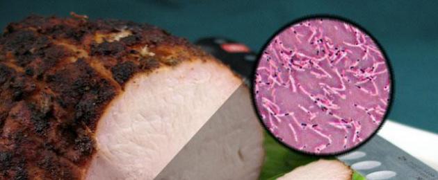
Spore bacteria are more resistant to freezing, drying, long-term or short-term boiling, and exposure to chemicals. There are vegetative forms of bacterial formation and sporulation. Examples of the latter: pathogens of diseases such as anthrax, botulism, tetanus and some types of saprophytic soil inhabitants that can be found in manure.
Germination
When the spore shell enters an environment favorable to it, it begins to swell. This process continues until the shell is completely destroyed. At the time when the membrane tissue ruptures, through this large burst the young cell enters the external environment.
In this way, the spore germinates in aerobic bacteria. Anaerobic bacteria do not lose their outer cell membrane during sporulation. The spore does not come into contact with the external environment; its contact occurs with the cell membrane. When favorable conditions arise, nutrients enter the cell through the cover. The spore begins to germinate.
Bacteria and products
Sporulation of bacteria is undesirable during processing and storage of certain products. If this process occurs, it will be difficult to fight microorganisms. To destroy spores in canned food, for example, the product must be sterilized, which will greatly reduce its quality. In order to preserve milk for a long time, it is sterilized, and this leads to the loss of its original properties and vitamin A. Note: the heating temperature during sterilization is 120 degrees.

During pasteurization, in order to preserve nutrients as much as possible, milk is heated to only 80-90 degrees. This affects the shelf life: milk quickly deteriorates, since during pasteurization the spores do not die, but on the contrary, they germinate and begin to multiply quickly, which is why the product deteriorates.
Controversy - form of resting gram-positive bacteria. Spores are formed under unfavorable conditions for the existence of bacteria (drying, nutrient deficiency, etc.). In this case, one spore is formed inside one bacterium. The formation of spores contributes to the preservation of the species and is not a method of reproduction. Spore-forming rod-shaped aerobic bacteria in which the spore size does not exceed the diameter of the cell are called bacilli. Spore-forming rod-shaped anaerobic bacteria in which the spore size exceeds the size of the bacterial cell are called clostridia.
Scheme of spore formation (according to G. Schlegel). A and B – septum formation. C and D – the spore protoplast is surrounded by the membrane of the mother cell. D – formation of the cortex and spore membranes. E – diagram of the structure of a mature spore: 1 – cytoplasm with nucleoid; 2 – CM spores; 3 – spore cell wall; 4 – cortex; 5 – inner shell of the spore; 6 – outer shell of the spore; 7 – exosporium.
Sporulation process(sporulation) goes through a number of stages. First, at one of the poles of the bacterial cell, the nucleoid condenses and separates due to the formation of a septum. Then the CPM begins to overgrow the formed protoplast of the spore and a fold appears, consisting of two layers of the CPM, later they merge, as a result of which the formed forespore is surrounded by a double shell. Between the membranes facing each other, the embryonic wall, the cortex, and the outer and inner membranes located outside the membranes are formed.
Spores are a peculiar form of resting firmicute bacteria, i.e. bacteria with a gram-positive type of cell wall structure. Spores are formed under unfavorable conditions for the existence of bacteria (drying, nutrient deficiency, etc. One spore (endospore) is formed inside the bacterial cell. The formation of spores contributes to the preservation of the species and is not a method of reproduction, like fungi. Spore-forming bacteria of the genus Bacillus have spores that do not exceed cell diameter. Bacteria in which the size of the spore exceeds the diameter of the cell are called clostridia, for example, bacteria of the genus Clostridium (lat. Clostridium - spindle). The spores are acid-fast, therefore they are stained red using the Aujeszky method or the Ziehl-Neelsen method, and the vegetative cell is stained red Blue colour.
The process of spore formation goes through a number of successive stages:
preparatory Metabolism changes, DNA replication is completed and condensation occurs. The cell contains two or more nucleoids, one of them is localized in the sporogenic zone, the rest are in the cytoplasm of the sporangium. At the same time, dipicolinic acid is synthesized;
prespore stage. On the side of the cytoplasmic membrane of the vegetative cell, an ingrowth of a double membrane, or septa, occurs, separating the nucleoid with an area of densified cytoplasm (sporogenic zone). As a result, a prospore is formed, surrounded by two membranes;
shell formation. First, a rudimentary peptidoglycan layer is formed between the membranes of the prospore, then a thick peptidoglycan layer of the cortex is deposited above it and the spore membrane is formed around its outer membrane;
spore maturation. The formation of all spore structures is completed, it becomes heat-resistant, acquires a characteristic shape and occupies a certain position in the cell.
15. Complex painting methods. Gram staining, its stages.
Consistently apply certain dyes, differing in chemical composition and color, mordants, alcohols, acids, etc. to the preparation. This makes it possible to identify cell structures and differentiate some types of microorganisms from others.
Gram stain.
1. Apply a carbolic alcohol solution of gentian violet to the fixed smear of the preparation through a strip of filter paper. After 1-2 minutes, remove it and wash off the dye.
2. Apply Lugol’s solution for 1-2 minutes
3. Decolorize with ethyl alcohol for 30-6 seconds until the violet streams of dye stop coming out.
4. Rinse with water.
5. Finish with an aqueous solution of fuchsin for 1-2 minutes, rinse, dry and microscope. Gram+: dark purple color. Gram -: red
16. Ziehl-Neelsen staining and its stages.
The Ziehl-Neelsen staining method is a method of staining microorganisms to identify acid-fast mycobacteria (pathogens of tuberculosis, mycobacteriosis, leprosy), actinomycetes and other acid-fast microorganisms. The acid resistance of microorganisms is due to the presence of lipids, waxes and hydroxy acids in their cells. Such microorganisms are poorly stained with diluted dye solutions. To facilitate the penetration of the dye into the cells of microorganisms, Zil's carbolic fuchsin applied to the preparation is heated over a burner flame. Colored microorganisms are not discolored by weak solutions of mineral acids and alcohol.
A smear fixed on a burner flame is stained for 3–5 minutes. with a solution of Ziehl's carbolic fuchsin or fuchsin-dyed paper, heated until vapor appears, but without bringing the dye to a boil.
Allow the preparation to cool, remove the paper, drain off the excess dye, and wash the preparation with water.
The colored preparation is decolorized with a 5% solution of sulfuric acid for 3–5 s or 96° ethyl alcohol containing 3% by volume hydrochloric acid, several times immersing the glass with a smear into a glass with hydrochloric acid alcohol.
After decolorization, the remaining acid is drained and the preparation is thoroughly washed with water.
Additional staining is done with Loeffler’s methylene blue for 3–5 minutes.
The colored preparation is washed with water, dried and microscopically examined.
When staining preparations, acid-fast bacteria are stained with fuchsin in a ruby-red color and are not discolored by acid.
Non-acid-fast bacteria, as well as tissue elements and leukocytes, become discolored under the influence of acid and acquire the color of an additional dye.
Controversy
One of the features of microorganisms is their ability to form spores. Spores are formed under unfavorable living conditions (drying, lack of nutrients, changes in pH, etc.), and only one spore is formed from one cell. Thus, the formation of spores is not associated with the process of reproduction, but is a kind of adaptation to surviving in unfavorable conditions. According to the accepted nomenclature spore-forming aerobes are called bacilli, A spore-forming anaerobes - clostridia.
Sporulation process passes series of stages, during which in a certain place the cell cytoplasm, nucleoid, ribosomes are concentrated, compacted, covered with a membrane, and then with a dense, poorly permeable multilayer membrane, including calcium salts of dipicolinic acid, which determines the heat resistance of spores. Spores can remain dormant for a long time, remaining viable. Thus, spores of pathogenic microorganisms (anthrax, tetanus, etc.) can persist in the soil for decades. When placed in a favorable environment, spores germinate very quickly - from 1 spore, 1 bacterial cell arises, which begins to multiply.
Sporulation is a species property of rods, and the shape and location of the developing spore in relation to the vegetative part of the cell is a differential diagnostic feature. Dispute form May be oval or round, location central(the causative agent of anthrax), subterminal- closer to the end of the stick (causative agents of gas gangrene, botulism) and terminal- at the end (the causative agent of tetanus).
In a mature spore, the following are distinguishable: a central, poorly stained area (sporoplasm), a two-layer CPM and a spore shell.
Sporoplasm (spore protoplast) includes cytoplasm, bacterial chromosome, protein synthesis systems and some others (for example, anaerobic energy production).
The spore shell is two-layered: the space between the layers is filled with glycopeptide polymers similar to peptidoglycans, forming a network structure (cortex) that is highly sensitive to lysozyme. The inner layer (spore wall) is formed by peptidoglycans, similar to those of the vegetative cell. The outer layer (the shell itself) is formed by keratin-like protein structures with low permeability.
The process of sporulation (spore formation) begins immediately after a nutrient deficiency occurs and lasts approximately 8 hours. No external sources of nutrition or energy are required. Sporulation is stimulated by the introduction of glucose, phosphorus and NH4 into the medium; inhibits the introduction of peptone, lactose, NaCl, CaCl 2 (in bacteria of the genus Bacillus - DL-alanine).
Sporulation is controlled by special genes. Their number varies among different species and can reach 70. Details of sporulation serve as species characteristics, but its fundamental principles are the same for all bacteria.
1.Preparatory stage sporulation is accompanied by a cessation of division and an increase in the number of lipid inclusions.
Forespore stage sporulation usually begins vigorously. An elliptical membrane appears in the cell, surrounding a section of cytoplasm with altered density and tinctorial properties. Such a formation is designated by the terms “prespore” or “primordial spore”.
1.The third stage of sporulation includes appearance of the shell(usually within 10 minutes after the formation of the forespore) and an even greater increase in the refractive index.
1.Maturation stage spores are accompanied by compaction and a decrease in the metabolic activity of the cell.
In Bacillus species, the diameter of the spore does not exceed the width of the cell, while in many Clostridium species it is significantly greater. In some cases, the cell acquires a very characteristic appearance, which is often a diagnostic sign. For example, terminally located spores in the causative agent of tetanus give the bacteria the appearance of “tennis rackets.”
Because the spores refract light strongly, then recognizing them under microscopy is not difficult. In controversial cases, they resort to special staining methods.
There are some types of bacteria that produce round or oval shaped bodies that are highly refractive to light. These formations are called endospores. Sporulation is one of the stages of the development cycle of certain microorganisms in response to the adverse effects of the external environment, developed during the process of evolution in the struggle to preserve the species. The lack of nutrients causes various reactions in some microorganisms that prepare the cell for a long period during which nutrients are not available. The transition to sporulation is observed when the nutrient substrate is depleted, with a lack of carbon, nitrogen or phosphorus, a change in the pH of the environment, etc. Sporulation is characteristic mainly of rod-shaped microorganisms (bacillus and clostridia, and is relatively rarely observed in cocci (Sarcina urea, Sarcina lutea) and convoluted forms (Desulfovibrio desulfuricans).
Sporulation occurs in the external environment, on nutrient media, and is not observed in human and animal tissues. The process of sporulation is divided into seven successive stages, characterized by various cytological changes (Fig. 12).
Preparatory stages(stages 0 and I). At these stages, there are still no morphologically visible changes in the cell, but the amount of water decreases and the cytoplasm becomes denser.
Prospore stage(stage II) is the first stage of sporulation, recognizable morphologically. It is characterized by the appearance of a prospore septum, which divides the cell into a small prospore and a large mother cell. This is a key stage of sporulation.
During stages of prospore absorption(stage III) spatial separation of a small prospore occurs, which passes into the cytoplasm of the mother cell. A double membrane structure is formed on the outside of the prospore.
Forespore stage characterized by the formation of a cortex (dense spore membrane) inside the membrane structure of the prospore (stage IV) and the condensation of proteins on its surface (stage V).
On maturation stages(stage VI) the spore coat develops further and becomes resistant to chemical agents and heat. The formed spore occupies approximately 1/10 of the mother cell.
The final stage is release of mature spores from the mother cell (stage VII). The process of spore formation occurs within 18-20 hours.
Due to the presence of a dense multilayer shell with a lamellar structure, a minimal amount of water and a high content of calcium, lipids and dipicolinic acid, the spores are highly resistant to environmental factors and disinfectants. They can withstand relatively high and low temperatures, prolonged drying, exposure to radiation, toxic substances, etc. They can survive for decades in unfavorable conditions.
Once in favorable conditions, the spores germinate and again turn into vegetative forms. The process of spore germination begins with the absorption of water. They swell and increase in size. A process appears from the shell at the pole, in the center, or between the pole and the center, from which the rod extends. The process of spore germination is much faster and takes 4 - 5 hours.
According to the nature of localization in the body of microorganisms, spores are located:
1. Central (anthrax bacillus, anthracoid bacillus, etc.).
2. Subterminal - closer to the end (causative agent of botulism, etc.).
3. Terminal - at the end of the stick (tetanus agent).
In certain types of spore-forming microorganisms, the diameter of the spores exceeds the diameter of the bacterial cell. If the spores are localized subterminally, such bacteria take on a spindle shape. These include butyric acid fermentation clostridia. In some clostridia, for example, in the causative agent of tetanus, the spores are located terminally, their cell resembles a drumstick (Fig. 13).

Rice. 13. Shapes and location of spores in bacilli.
The ability to form spores is used in the taxonomy of microbes, as well as when choosing methods for disinfecting objects, premises, food products, and various products.
- In contact with 0
- Google+ 0
- OK 0
- Facebook 0