Bone
The bone tissue that forms the bones of the skeleton is very strong. It maintains the shape of the body (constitution) and protects the organs located in the cranium, chest and pelvic cavities, participates in mineral metabolism. The tissue consists of cells (osteocytes) and an intercellular substance in which nutrient channels with vessels are located. The intercellular substance contains up to 70% of mineral salts (calcium, phosphorus and magnesium).
In its development, bone tissue goes through fibrous and lamellar stages. In various parts of the bone, it is organized in the form of a compact or spongy bone substance.
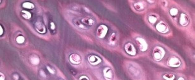
Cartilage tissue consists of cells (chondrocytes) and intercellular substance (cartilaginous matrix), which is characterized by increased elasticity. It performs a supporting function, as it forms the bulk of the cartilage.
There are three types of cartilage tissue: hyaline, which is part of the cartilage of the trachea, bronchi, ends of the ribs, articular surfaces of bones; elastic, forming the auricle and epiglottis; fibrous, located in the intervertebral discs and joints of the pubic bones.

Adipose tissue is similar to loose connective tissue. The cells are large and filled with fat. Adipose tissue performs nutritional, shaping and thermoregulatory functions. Adipose tissue is divided into two types: white and brown. In humans, white adipose tissue predominates, part of it surrounds the organs, maintaining their position in the human body and other functions. The amount of brown adipose tissue in humans is small (it is present mainly in a newborn child). The main function of brown adipose tissue is heat production. Brown adipose tissue maintains the body temperature of animals during hibernation and the temperature of newborns.

Muscle cells are called muscle fibers because they are constantly elongated in one direction.
The classification of muscle tissues is carried out on the basis of the structure of the tissue (histologically): by the presence or absence of transverse striation, and on the basis of the mechanism of contraction - voluntary (as in skeletal muscle) or involuntary (smooth or cardiac muscle).
Muscle tissue has excitability and the ability to actively contract under the influence of nervous system and some substances. Microscopic differences make it possible to distinguish two types of this tissue - smooth (non-striated) and striated (striated).
smooth muscle tissue has a cellular structure. It forms the muscular membranes of the walls of internal organs (intestines, uterus, bladder, etc.), blood and lymphatic vessels; its contraction occurs involuntarily.

striated muscle tissue consists of muscle fibers, each of which is represented by many thousands of cells, merged, in addition to their nuclei, into one structure. It forms skeletal muscles. We can shorten them as we wish.

A variety of striated muscle tissue is the heart muscle, which has unique abilities.

During life (about 70 years), the heart muscle contracts more than 2.5 million times. No other fabric has such strength potential. Cardiac muscle tissue has a transverse striation. However, unlike skeletal muscle, there are special areas where the muscle fibers meet. Due to this structure, the contraction of one fiber is quickly transmitted to neighboring ones.
This ensures the simultaneous contraction of large sections of the heart muscle.
nervous tissueNervous tissue consists of two types of cells: nervous (neurons) and glial. Glial cells are closely adjacent to the neuron, performing supporting, nutritional, secretory and protective functions.

The neuron is the basic structural and functional unit of the nervous tissue. Its main feature is the ability to generate nerve impulses and transmit excitation to other neurons or muscle and glandular cells of the working organs. Neurons may consist of a body and processes. Nerve cells are designed to conduct nerve impulses. Having received information on one part of the surface, the neuron very quickly transmits it to another part of its surface. Since the processes of a neuron are very long, information is transmitted over long distances. Most neurons have processes of two types: short, thick, branching near the body - dendrites and long (up to 1.5 m), thin and branching only at the very end - axons. Axons form nerve fibers.
The images below will take you on a journey through your body, from your head to your intestines and pelvic organs. You'll see what normal cells look like and what happens to them when cancer strikes them, and you'll also get a visual representation of how, say, the first meeting of an egg and sperm occurs.
Almost all of the images presented here were taken with a scanning electron microscope (SEM). The beam of electrons emitted by such a device interacts with the atoms of the desired object, resulting in 3D images of the highest resolution. A magnification of 250,000 times allows you to see details of 1-5 nanometers in size (that is, billionths of a meter).
Max Knoll obtained the first SEM image in 1935, and already in 1965 the Cambridge Tool Company offered its Stereoscan to DuPont. Now such devices are widely used in research centers.
The images below will take you on a journey through your body, from your head to your intestines and pelvic organs. You'll see what normal cells look like and what happens to them when cancer strikes them, and you'll also get a visual representation of how, say, the first meeting of an egg and sperm occurs.
Here is depicted, one might say, the basis of your blood - red blood cells (RBC). These pretty biconcave cells are responsible for carrying oxygen throughout the body. Usually in one cubic millimeter of blood there are 4-5 million such cells in women and 5-6 million in men. People living in the highlands, where there is a lack of oxygen, have even more red cells.

To avoid this kind of hair splitting that is invisible to the ordinary eye, you need to cut your hair regularly and use good shampoos and conditioners.

Of the 100 billion neurons in your brain, Purkinje cells are among the largest. Among other things, they are responsible in the cerebellar cortex for motor coordination. They are detrimental to both alcohol and lithium poisoning, as well as autoimmune diseases, genetic abnormalities (including autism), as well as neurodegenerative diseases (Alzheimer's, Parkinson's, multiple sclerosis, etc.).

This is what stereocilia look like sensitive elements vestibular apparatus inside your ear. Capturing sound vibrations, they control the response mechanical movements and actions.

Shown here are retinal blood vessels emerging from a black-stained optic disc. This disk is a "blind spot" because there are no light receptors in this area of the retina.

There are about 10,000 taste buds on the human tongue, which help to determine the taste of salty, sour, bitter, sweet and spicy.

In order to avoid such layers similar to non-threshed spikelets on the teeth, it is advisable to brush your teeth more often.

Remember how beautiful healthy red blood cells looked. Now look at what they become in the web of a deadly blood clot. In the very center is a white blood cell (leukocyte).

Here is a view of your lung from the inside. Empty cavities are alveoli where oxygen is exchanged for carbon dioxide.

And now take a look at how the lungs deformed by cancer differ from healthy ones in the previous picture.

The villi of the small intestine increase its area, which contributes to better absorption of food. These are outgrowths of irregular cylindrical shape up to 1.2 mm high. The basis of the villi is loose connective tissue. In the center, like a rod, there is a wide lymphatic capillary, or milky sinus, and on the sides of it there are blood vessels and capillaries. Through the lactiferous sinus, fats enter the lymph, and then into the blood, and proteins and carbohydrates enter the bloodstream through the blood capillaries of the villi. On closer examination, you can see food residues in the grooves.

Here you see a human egg. The egg is covered with a glycoprotein coat (zona pellicuda), which not only protects it, but also helps to capture and hold the sperm. Two coronal cells are attached to the shell.

The picture captures the moment when several spermatozoa are trying to fertilize the egg.

It looks like a war of the worlds, but in fact, you have an egg in front of you 5 days after fertilization. Some spermatozoa are still held on its surface. The image was taken using a confocal (confocal) microscope. The egg and sperm nuclei are purple, while the sperm flagella are green. The blue areas are nexuses, intercellular gap junctions that communicate between cells.

You are present at the beginning of a new life cycle. A six-day-old human embryo is implanted in the endometrium, the lining of the uterine cavity. We wish him good luck!
It's hard to even imagine that a human body can look like this...
First seen on the pages school textbook in anatomy, what organs the human body consists of, each of us has made a small or big discovery for himself. And certainly, since then, no one else has been taken lightly to their “well-coordinated mechanism”, where even the smallest detail is important ...
But it seems that the time has come to make a new step in self-knowledge and look even deeper - into a microscope!
Get ready, we found 23 images of human organs taken using electron microscopy, from which everything inside you will “turn over”!
1. You won't believe it, but this is what your eye looks like when you get as close as possible!
2. And like this - the base of the nail, which you are now tapping on the keyboard ...

3. Are you breathing calmly? So it's thanks to the cells of the lungs!

4. And this is how lung cells look even closer!


6. Well, if you are now dizzy, then be sure that the balancing stones in the inner ear may well be the reason for this!

7. Incredible moment - this is what an artery and blood look like!

8. And you, too, were haunted by these red blood cells, which were told in the lessons? So look at them...

9. Calmness, only calmness - it's just a blood clot!

10. Another unique shot - red blood cells come out of a broken capillary.

11. Well, it's time to see how the "critical days" begin!

12. And this is what fat cells look like after you went on a diet and lost a couple of kg!

13. And if you suddenly doubt whether to treat the split ends of the hair or not, then look how bad they are at this moment ...

14. And this is how our hair looks at the root!

15. And after this photo, you will no longer go to bed until you remove your eye makeup - these are our cilia!

16. Want to show your tongue? And today you cleaned it during the morning procedures?

17. By the way, sweat on the surface from the pores looks almost as disgusting as it “smells” ...

18. And this is the gastric mucosa. But you certainly imagined it quite differently!

19. Haven't bought a hand cream for a long time? But in vain - the fingertips definitely need it!

20. Well, it's time to finally find out what it looks like at the maximum approximation ... sperm!

21. And this is how it looks in the seminal canals? Suddenly?

22. And this is how each of us was on the sixth day of life in the womb! Isn't that happiness?

23. But happiness from the closest distance looks exactly like this - crystals of the hormone serotonin!

With in-depth studies of microscopy, it becomes important for novice biologists and physicians to study histological samples. They are prepared using a special technology with the dissection of biological tissue into thin sections using a microtome. We will talk about this briefly in this review using the example of a study of the brain under microscope. We will need a binocular or trinocular model with a lower illuminator providing a method of observation in transmitted light (bright field).
Brain is located in the brain region of the skull (bone part of the head) of humans and vertebrates, and is the main organ of the central nervous system. In this center of control of the activity of a living organism, due to the synoptic transmission of nerve impulses, many electrically excitable neurons are combined.
Currently, the brain is not fully understood, many aspects remain unclear, despite the large number of laboratories in anatomy and architectonics, and the huge amount of work done by scientists around the world. It is known that in humans its mass is equal to an average of two percent of the total body weight. It has a complex structure and wide functionality.

Tissues that can be seen in a microslide of the brain under the microscope:
- Connective fibrous fibrous. Forms hard, arachnoid and pia mater. The main cells in its composition are: fibroblasts, synthesizing components of the intercellular substance;
- Cerebrospinal fluid (called "CSF"), which performs protective functions and continuously circulates in the lateral, third and fourth ventricles (cavities). It also ensures the maintenance of intracranial pressure favorable for life. It is produced by vascular plexuses - formations that, with a 1000-fold increase, are distinguishable as villi;
- Nerve fibers are visible processes of neurons covered with glia;
- Glial cells.
- A network of elastic blood vessels made up of myocytes.
Without special medical equipment, it will not be possible to prepare a micropreparation on your own, in this case it is recommended to use already ready sample included in the "Anatomy and Physiology" kit (Micromed or Levenhuk).

Stages of creating a microsample in a pathoanatomical laboratory:
- Taking biomaterial for diagnosis by a surgeon or pathologist;
- Fixation in formalin or alcohol solution.
- Hematoxylin-eosin staining
- Freeze. Deep cooling contributes to the hardening required for cutting by microtomy;
- Installation between the slide and cover slip.
The microsample is placed in the slider or under the metal clips of the microscope stage. Then it is centered in such a way that light radiation penetrates the preparation from below, passing through the cellular structure upwards towards the optical system. The condenser is adjusted for maximum light transmission. Initially, a “search” lens of the minimum magnification is selected on the revolver, then the degree of approximation is increased step by step to 400x and 1000x.
results research activities are fixed in the form of photographs - for this, a digital camera is inserted into one of the eyepiece tubes of the visual attachment and connected to a computer. Photographing is carried out by software.
Topic : The study of the structure of cells and body tissues under a microscope .
1. To study the cell and tissues of the human body. 2. Develop skills of observation, comparison, working with a microscope.
3. Education of a culture of behavior.
Method: visual, verbal, practical.
Lesson type: Combined.
Equipment: Microscopes, prepared slides.
During the classes:
1.org moment.
2. Control homework.
Test check.(1-5)
Checking the execution of the table in the notebook. Page: 28.
3. Studying a new topic.
Laboratory work № 1.
.
Purpose: Acquaintance with the structure of the cell and tissues of the human body under a microscope.
Operating procedure:
1. Consider finished preparations of the structure of cells of different tissues. Find structural elements (membrane, cytoplasm, nucleus)
2 Draw the examined cells indicating the organelles seen under the microscope.
3. Examine tissue under a microscope:
A) striated muscle
b) striated cardiac;
B) smooth muscle
D) epithelial (various epithelium)
D) bone.
E) nervous.
Draw conclusions:
1. What are the similarities and differences in the structure of these tissues.
2. Locate the structural parts of these tissues.
3. Determine whether the cells are located in the same way in the tissues?
4. Draw the examined fabrics. Sign them. Compare with the pattern in the textbook.
Complete the table in your notebook.
Fabrics and their functions
Fabric name
structure
Location in the body
striated
Smooth muscle
Connective
epithelial
Homework: §7 and 8.
Grade 8 performs laboratory work No. 1.
Topic: Pancreas, adrenal glands, gonads.
1. To study the function of mixed glands.
2. Develop knowledge in the field of physiology.
Lesson type: Combined.
Equipment. Tables.
During the classes:
1.org moment.
2. Homework control.
Performing exercises No. 1-No. 3.
3. Studying a new topic.
Pancreas:
1. Structure: head, body, tail.
2.Function: forms digestive juice with enzymes.
(external secretory function)
intrasecretory cells produce the hormones insulin and glucagon that regulate carbohydrate metabolism.
adrenal glands
1.building
2.function.
sex glands:
1.Building
2.Function.
4. Fixing.
Working with terms.
5. Evaluation.
:.Homework.§12-13.
Topic: Structure and functions of the nervous system.
1. Study the structure of a neuron
2. Develop interest in anatomy and physiology.
Lesson type: Combined.
Equipment: Table.
During the classes:
1.org moment.
2. Homework control.
Execution of the table p.43. Poll on ZhVS.
3. Studying a new topic.
Neuron
2. Dendrites
4. Receptors.
At the junctions of dendrites, synapses.
Transmission of signals through synapses is carried out with the help of neurotransmitters.
4. Fixing.
Reading text.
Draw a neuron from memory.
5. Evaluation.
6. Homework. §14.
Topic : Reflex. Reflex arc.
1. To study the function of the reflex arc using the knee jerk as an example.
2. Develop observation, attentiveness.
3. Education of industriousness.
Method: verbal, visual, practical.
Lesson type: Combined.
Equipment:
Rubber mallet.
During the classes:
1.org moment.
2. Homework control.
Draw and indicate parts of a neuron.
3. Studying a new topic.
Reflex is the body's response to irritation.
The reflex arc is the path along which the nerve impulse travels.
Sensory receptors conduct impulses to the nervous system. in a centripetal way.
Motor receptors respond to the reflex.
By centrifugal way.
4. Fixing.
Lab #2
"Knee Reflex Study"
Draw a reflex arc. Highlight parts with colored markers.

The work is carried out according to the plan, and submitted for verification.
5. Homework: §14.
Lesson number 14.
Topic : The structure and function of the spinal cord.
1. Study the structure of the spinal cord.
2. Develop research skills.
3. Education of scientific outlook.
Method: Verbal, visual, practical.
Lesson type: Combined.
Equipment: tables. Diagram of the spinal cord.
During the classes:
1.org moment.
2. Homework control.
Specify the parts of the knee jerk. Fill in the table. Poll on questions.
3. Studying a new topic.
Nervous system
Central peripheral.
The structure of the spinal cord.
The brain is located in the nutria of the spinal column. A cord with a diameter of 1 cm is white. The spinal cord consists of white and gray matter. (Find the characteristic in the text)
The spinal cord consists of 31 segments. The anterior and posterior roots form mixed nerves.
The anterior make up the motor fibers;
The rear homies make up the sensitive fibers.
Spinal Cord Function:
1.Reflex
2. Conductor.
4. Fixing.
Fill in the table.
5. Homework: §15.
Lesson number 15.
Topic: The structure and functions of the brain. Large hemispheres, hygiene of the nervous system.
1 To study the main parts of the brain and their function.
2. Develop knowledge of the anatomy and physiology of the body.
3. Education of scientific outlook.
Method: verbal, visual, practical.
Lesson type: combined.
Equipment: tables, layout.
During the classes:
1.org moment.
2. Homework control:
Checking the filling of tables. pp. 49-50.
Run exercise No. 1. and No. 2 in workbook.
Exploring a new topic .
1. Brain
Structure function
medulla oblongata sucking, swallowing, coughing, sneezing.
The bridge connects the oblong with the middle
Midbrain reaction to light, sound, muscle tone
The diencephalon conducts impulses to the BP cortex. ,walking,
Swimming, regulates the exchange of v-in, needs
Leaning water and food.
Cerebellum movement coordination.
2. Large hemispheres:
Structure function
Formed by gray bark.; In the left p.sh. in right-handers, and in the right of the lion
Area 2200-2500cm 3 necks - auditory and motor center of speech
Furrows: large divided into: and letters;
The frontal and parietal are the deepest; Right p.sh. creative thinking, Musical creativity,
Occipital, temporal,
Formed by the white in-tion;
Sensory areas located
(sensitive)
Temporal - auditory;
Olfactory and gustatory - on the border
Parietal and temporal;
3. Hygiene of the nervous system:
School hygiene; Alcohol,
Their effect on the nervous system?
4. Fixing.
Work with text §16.17.Workbook. Exercise No. 1, No. 2.
5. Evaluation.
6. Homework §16 -17..
Lesson number 1. 3.09.12
Topic : Introduction. Goals and objectives of the subject.
“The health of the people is above all,
The riches of the earth will not replace him.
Health can not be bought, no one will sell.
Take care of him like a heart, like an eye.
1. Bring to students the goals and objectives of the subject ..
2. Develop knowledge of human anatomy and physiology.
3. Education of occupational health.
Method: verbal, visual, practical.
Lesson Type: Introductory.
Equipment. Tables.layouts.
During the classes:
1.org moment.
2. Homework control.
Test check. (Zero slice of knowledge)
3. Studying a new topic.
1. Acquaintance with the structure of the textbook.
Registration laboratory notebooks,
Reading the introductory part.
4. Fixing. Task number 1. Name the sciences that study the structure and functions human body.
1….. 3………
2…… 4………. 5………..
5. Homework: §1.
Lesson No. 2 7. 09.12.
Topic : Methods for studying the structure and functions of the body.
1. Study what anatomy and physiology studies ..
2. Develop the concepts of anatomy and physiology.
Method: Verbal, visual, practical.
Lesson Type: Educational.
Equipment.
During the classes.
1.org moment.
2. Homework control.
Which treatise wrote Charles Darwin and what is its significance.
What branches of science does a person study?
What are the goals and objectives of humanism?
3. Studying a new topic.
The class is divided into two teams.
1 team.
Task number 1. What does anatomy study?
No. 2. What techniques and methods does anatomy use in the study of a person?
2.team.
Task number 1. What does physiology study?
No. 2. What techniques and methods does physiology use in the study of man?
Homework: §2.
Lesson number 3. 10.09.12
Topic: Cell structure human body.
1. To study the types of cell shapes, sizes and composition of cells.
2. Develop an understanding of cell diversity.
3. Education of scientific outlook.
Method: verbal, visual, practical.
Lesson type: educational.
Equipment. Tables.
During the classes.
1.org moment.
2. Homework control.
What methods and techniques are used in the study of physiological processes?
(microscope, kymograph, electromagnetic tuning fork. Electrocardiograph..).
What help do you use chemical methods physiology?
3. Learning a new topic:
1. Reading a paragraph with marks.
What do I know.
What did you find out.
I want to know.
4. Fixing.
What are the types of cells in the human body?
What is the role of the kernel?
How do you understand the expression of a pore in a membrane?
5.Homework§3
Lesson number 4. 09/14/12
Topic : Cell organelles, chemical composition cells.
1. to study organelles and their chem. compound.
2. To develop knowledge in the field of cytology.
3. Education of scientific outlook.
Method: verbal, visual, practical.
Equipment. Table. electronic textbook.
During the classes:
1.org moment.
2 Control of homework.
What is the structure of a cell?
How is the plasma membrane different from the cell wall?
What causes the acceleration and deceleration of the movement of the cytoplasm?
3. Studying a new topic.
cell organelles.
Using the poster, list the organelles of the cell.
EPS - Smooth and rough;
Ribosomes;
Mitochondria;
Lysosomes;
Golgi apparatus;
Centrioles.
Differences between plant and animal cells.:
Animals have centrioles; Plants have plastids
In vegetable there is cellulose;
There are large vacuoles.
Vital properties of the cell:
1. Metabolism;
Irritability;
Growth and development;
Reproduction.
4. Fixing.
Exercise number 2. Set a match.
Add suggestions.
The main structural and functional element of our body is .... (cell)
There is a nucleus in every cell, except ...... (erythrocytes)
5. Evaluation.
6. Homework: §4.
Lesson number 5. 09/17/12
Topic : The study of the structure of cells and tissues of the human body under a microscope.
1. Using laboratory work to study the structure of the cell.
2. Develop skills to observe and compare.
3. Education of scientific outlook.
Method: verbal, visual, practical.
Lesson type: combined.
During the classes:
1.org moment.
2. Homework control.
Check exercise number 2; No. 3.
Survey on questions. (Levels A, B, C)
3. Studying a new topic.
Laboratory work number 1.
The study of the structure of cells and tissues of the human body under a microscope.
Complete the table in your notebook.
Fabric name
Structure
location in the body.
striated
Smooth muscle
Connective.
epithelial,
Draw the considered fabrics.
4. Evaluation.
5.Home building§§7.8.
Lesson number 6. 09/21/12
Topic: Human body tissues, properties and functions.
1. to study human tissues.
3. Education of scientific outlook.
Method: verbal, visual, practical.
Lesson Type: Educational.
During the classes:
1.org moment.
2. Homework control.
Retell the work done.
3. Studying a new topic.
Epithelial. Connective Muscular. Nervous.
4. Fixing.
Exercise number 3. Distribute the signs and functions of tissues in the appropriate squares, first write the names of the tissues.
fabrics
epithelial
connective
muscular
5. Evaluation.
6. Homework. §8.
Lesson number 7. 09/24/12
Topic: Organs and organ systems.
1. To study the systems of organs and the organs that form them.
2. Develop skills to memorize drawings.
3. Education of scientific outlook.
Method: Verbal, visual, practical.
Lesson Type6
Combined.
During the classes.
1.org moment.
2. Homework.
Check Exercise No. 1 - No. 3.
Poll for questions.
3. Studying a new topic.
Using figure No. 14 of the textbook for memorization and Exercise No. 3 in the workbook for consolidation.
Repeat On the board a cluster of systems..
4. Evaluation.
5. Homework: § 9. Exercise No. 1-No. 3.
Lesson number 8. 09/28/12
Topic : The integrity of the human body.
1. To study the function of homeostasis.
2. Develop knowledge of cytology.
3. Education of labor discipline.
Method: Verbal, visual, practical.
Lesson Type: Educational.
Equipment.
During the classes:
1.org moment.
2. Homework control.
Name the organs of the respiratory system?
Task No. 1. Add suggestions.
The liver belongs to the (digestive) ... system.
The heart belongs to the (circulatory) system.
The paired organs that make urine are called the kidneys.
The excretory system includes (kidneys, ureters, urinary bladder)
3. Learning a new topic.
The integrity of the human body is formed by: blood, lymph, intercellular fluid.
Reading with notes.
Fill the table.
New information.
I want to know.
4. Fixing.
5. Homework: §10.
Lesson number 9. 01.10.12
Topic: Endocrine glands. Hormones. Pituitary.
1. To study the significance of the endocrine glands.
2. Develop an interest in biology.
3. Education of scientific outlook.
Method: verbal, visual, practical.
Lesson type, Combined.
Equipment: Tables, ZhVS.
During the classes.
1.org moment.
2. Homework control.
What is homeostasis disrupted?
3. Studying a new topic.
Humoral regulation of the body is carried out by biologically active substances.
The pituitary gland produces hormones.
In the body, the glands are divided into three groups.
1. Glands of internal secretion 2. Glands of mixed secretion.
3. Glands of external secretion.
The class is divided into three groups. Complete assignments and prepare presentations. .
4. Fixing.
Pituitary
Structure function.
5. Evaluation.
6. Homework: § 11
Lesson number 10. 0 8.10.12
Topic: Thyroid, parathyroid gland.
1. To study the structure and function of the glands.
2. Develop cognitive interest.
3. Education of the scientific worldview.
Method: Verbal, visual, practical.
Lesson type: educational.
Equipment. Table.
During the classes:
1.org moment.
2. Homework control.
Exercise No. 1- No. 4.
3. Studying a new topic.
Thyroid
Structure function
Parathyroid gland
Structure function.
4. Evaluation.
6. Homework: §12.
Lesson number 16.
Topic: Autonomic nervous system and its divisions.
1. Study the sympathetic and parasympathetic nervous system.
2. Develop skills in working with a textbook.
3. Education of labor discipline.
Method: Verbal, visual, practical. Lesson Type: Educational. Equipment: Table.
During the classes:
1.org moment.
2. Homework control.
Fill the table.
Functions of the sensory areas of the cerebral cortex.
The name of the zone.
Its functions
1. Zone of visual perceptions.
2. Hearing zone.
3. Centers of smell and taste.
4. Zones of skin and muscle sensitivity.
5. motor zone.
6. Associative zones.
3. Studying a new topic.
vegetative system.
sympathetic Parasympathetic.
(Using tutorial create a cluster).
1.increases blood pressure
2.dilates pupils
3. enhances heat dissipation.
4. speeds up the heartbeat.
5. inhibits the activity of the zh.k.t.
6 Relaxes the smooth muscle tissue of the bladder.
4. Fixing.
Remember! The autonomic nervous system. Sympathetic and parasympathetic division.
5. Evaluation.
6.Homework: §18.table.
Topic: Sense organs and their meaning.
1. Study the function of the analyzers.
2. Develop observation.
3. Education of labor discipline.
Method: verbal, visual, practical.
Lesson type: Combined.
Equipment: table.
Lesson progress 6
1.org moment.
2. Homework control.
Fill in the table.
Functions of the divisions of the autonomic nervous system
Organs and systems
Actions of the sympathetic department
Actions of the parasympathetic division
Blood vessels
Digestive system
urinary system
3. Studying a new topic.
Sense Organs: Analyzers: Visual
Auditory
Touch
Smell
4. Fixing.
1. Reading a paragraph.
2. Work with the table.
Set a match.
1.Receptor
2. Sensitive nerves.
3. Zones of the cerebral cortex
A) transmission of impulse to the working body.
C) the perception of irritation.
C) transmission of excitation from receptors.
D) excitation transfer
D) the formation of sensations.
5. Evaluation.
6. Homework: §19.
- In contact with 0
- Google+ 0
- OK 0
- Facebook 0