"Introduction to general biology and ecology. Grade 9. A.A. Kamensky (gdz)
The structure and functions of mitochondria, plastids and lysoso m
Question 1. Where is the lysosome formed?
Lysosomes- single membrane organelles general type. Membrane vesicles containing degrading enzymes.
Lysosome classification:
primary - lysosomes, which contain only the active enzyme (eg acid phosphatase);
secondary - these are primary lysosomes along with the substance that is digested (autophagosomes - break down the internal parts of the cell that have performed their functions; heterophagosomes - break down substances and structures that have entered the cell). Residual bodies are secondary lysosomes containing undigested material.
Lysosomes are formed in the Golgi apparatus, where they enter and where enzymes accumulate.
Question 2. What is the function of mitochondria?
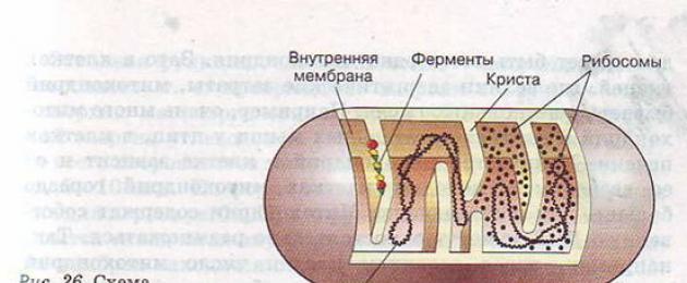
Mitochondria- organelles of a general type, having a two-membrane structure. The outer membrane is smooth, the inner one forms outgrowths of various shapes - cristae. In the mitochondrial matrix (semi-liquid substance), between the cristae, there are enzymes, ribosomes, DNA, RNA, which are involved in the synthesis of mitochondrial proteins. Mushroom bodies are visible on the inner membrane - ATP-somes, which are enzymes that form ATP molecules.
Functions:
1) ATP synthesis;
2) participate in carbohydrate and nitrogen metabolism: a) on the outer membrane and nearby in the hyaloplasm goes anaerobic oxidation(glycolysis); b) on the inner membrane - cristae - there are processes associated with the oxidative cycle of tricarboxylic acids and the respiratory chain of electron transport, i.e. cellular respiration, as a result of which ATP is synthesized;
3) have their own DNA, RNA and ribosomes, i.e. they can synthesize proteins themselves;
4) synthesis of some steroid hormones.
Question 3. What types of plastids do you know?
plastids-two membrane organelles plant cells general type, are divided into three types:
a) leukoplasts - microscopic organelles having a two-membrane structure. The inner membrane forms 2-3 outgrowths. The shape is rounded. Colorless. located in plant organs inaccessible to sunlight(for example, in rhizomes, tubers). When exposed to light, chlorophyll is formed in them. Functions: the center of accumulation of starch and other substances. In the light they are converted into chloroplasts.
b) chromoplasts - microscopic organelles having a two-membrane structure. Actually chromoplasts have a spherical shape, and those formed from chloroplasts take the form of carotenoid crystals, typical for this plant species. Coloring red, orange, yellow. They are located mainly in the fruits and petals of flowers, which gives these plant organs a corresponding bright color. Functions: contain red, orange and yellow pigments (carotenoids). A lot of ripe fruits of tomatoes and some algae; color the corolla of flowers.
c) chloroplasts are microscopic organelles having a two-membrane structure. The outer membrane is smooth. The inner membrane forms a system of two-layer plates - thylakoids of the stroma and thylakoids of the gran. The thylakoid is a flattened sac. A grana is a stack of thylakoids. Pigments - chlorophyll and carotenoids - are concentrated in the membranes of thylakoid gran between the layers of protein and lipid molecules. The protein-lipid matrix contains its own ribosomes, DNA, RNA, starch grains. The shape of chloroplasts is lenticular. The coloring is green. Functions: photosynthetic, contain chlorophyll. On the grana is the light phase of photosynthesis, in the stroma - the dark phase.
plastids- organelles that have their own genetic information and synthesize their own proteins.
Question 4. How does each type of plastid differ from another?
plastids different types differ from each other by the presence or absence of certain pigments. Leukoplasts lack pigments, chloroplasts contain green pigments, and chromoplasts contain red, orange, yellow, and violet pigments.
Question 5. Why are the grana in the chloroplast staggered?
Grana in chloroplasts are staggered so as not to block each other from sunlight. Sunlight should illuminate each grain well, then photosynthesis will proceed more intensively.
Question 6. What will happen if the lysosome in one of the cells is suddenly destroyed?
With a sudden rupture of the membrane surrounding the lysosome, the enzymes contained in it enter the cytoplasm and gradually destroy the entire cell. Cytolysis occurs - the destruction of cells by their complete or partial dissolution both under normal conditions (for example, during metamorphosis), and with the penetration of pathogens, malnutrition, lack and excess of oxygen, improper use of antibiotics and under the action of toxic substances (pathological lysis).
Question 7. What are the similarities between mitochondria and plastids?
The morphological and functional organization of mitochondria and chloroplasts has the following common features:
Mitochondria and plastids have a two-membrane structure.
Chloroplast ribosomes, like mitochondrial ribosomes, synthesize proteins.
Chloroplasts, like mitochondria, reproduce by fission.
Both in mitochondria and in chloroplasts, ATP is synthesized (in mitochondria - during the breakdown of proteins, lipids and carbohydrates, and in chloroplasts - due to the conversion of solar energy into chemical energy).
The main characteristic that unites these organelles is that they have their own genetic information and synthesize their own proteins.
1. What is the structure and function of ATP?
Adenosine triphosphate (ATP) is a nucleotide consisting of a nitrogenous base adenine, a ribose carbohydrate and three phosphoric acid residues.
ATP is a universal source of energy for all reactions occurring in the cell.
2. What types of plastids do you know?
3. What methods of cell movement do you know?
1. Amoeboid movement.
2. Movement with the help of flagella and cilia.
3. Movement with the help of muscles.
4. In what form does the cell store nutrients?
in the form of lipids and glycogen.
Questions
1. What is the function of mitochondria?
The function of mitochondria is the synthesis of ATP.
2. What types of plastids do you know?
Plastids are divided into leukoplasts, chloroplasts, and chromoplasts based on color.
3. How does each type of plastid differ from another?
Leucoplasts - unstained plastids, as a rule, perform a storage function. In the leukoplasts of potato tubers, starch accumulates. Leucoplasts higher plants can transform into chloroplasts or chromoplasts.
Chromoplasts are plastids that are colored yellow, red, or orange. The coloration of chromoplasts is associated with the accumulation of carotenoids in them. Chromoplasts determine the color of autumn leaves, flower petals, root crops, and ripe fruits.
Chloroplasts are plastids that carry photosynthetic pigments - chlorophylls. They have a green color in higher plants, char and green algae.
4. Why are the grana in the chloroplast staggered?
The grains are staggered so that the light of the sun can reach each of them.
5. What are the similarities between mitochondria and plastids?
Like mitochondria, plastids contain their own DNA molecules. Therefore, they are also able to reproduce independently, independently of cell separation.
6. What are the functions of the cell center?
The cell center plays an important role in the formation of the internal skeleton of the cell - the cytoskeleton. Numerous microtubules diverge from the region of the cell center, supporting the shape of the cell and playing the role of a kind of rails for the movement of organelles through the cytoplasm.
The role of the cell center is great in cell division, when the centrioles diverge to the poles of the dividing cell and form the spindle of division.
7. Give examples of cellular inclusions.
These can be small drops of fat, starch or glycogen granules, less often - protein granules, salt crystals.
Tasks
Comparing the structure and functions of mitochondria and plastids. What are their similarities and differences?
Similarity:
● Two-membrane organelles. The outer membrane is smooth, and the inner one forms numerous invaginations that serve to increase the surface area. There is an intermembrane space between the membranes.
● Have their own circular DNA molecules, all types of RNA and ribosomes.
● Able to grow and reproduce by division.
● They carry out the synthesis of ATP.
Differences:
● Invaginations of the inner membrane of mitochondria (cristae) look like folds or ridges, and invaginations of the inner membrane of chloroplasts form closed disc-shaped structures (thylakoids) collected in stacks (granas).
● Mitochondria contain enzymes involved in the process of cellular respiration. The inner membrane of chloroplasts contains photosynthetic pigments and enzymes involved in the conversion of light energy.
● The main function of mitochondria is the synthesis of ATP. The main function of chloroplasts is photosynthesis.
Lysosomes. Mitochondria. plastids
1. What is the structure and functions ATP?
2. What types of plastids do you know?
When various nutrients enter the cell through phagocytosis or pinocytosis, they must be digested. Wherein squirrels must break down to individual amino acids, polysaccharides - to glucose or fructose molecules, lipids- to glycerol and fatty acids. In order for intracellular digestion to become possible, the phagocytic or pinocytic vesicle must fuse with the lysosome (Fig. 25). Lysosome is a small vesicle with a diameter of only 0.5-1.0 microns, containing a large set of enzymes that can destroy food substances. One lysosome can contain 30-50 different enzymes.

Subject: Lysosomes. Mitochondria. plastids
Target: to introduce students to the structure and functions of lysosomes, mitochondria and plastids.
During the classes
I . Organizing moment of the lesson
II . Repetition and consolidation of material
1. Structure and functions of the endoplasmic reticulum. The structure and functions of the Golgi complex.
(Student answers at the blackboard.)
2.
Why is the Golgi apparatus absent in erythrocytes?
What is the function of ribosomes? Why are most ribosomes located on channels in the endoplasmic reticulum?
What is the structure of ATP? Why is ATP called the universal source of energy for all reactions occurring in the cell?
3. "Silent" biological dictation
(The teacher points with a pointer to the table. Cell organelles, and students write down the names of organelles in notebooks)
1 - nucleus, 2 - nucleolus, 3 - ER, 4 - rough ER, 5 - cell membrane, 6 - cytoplasm, 7 - ribosome
III . Learning new material
The structure and functions of lysosomes.
Guys, let's remember in what ways various substances can penetrate inside the cell? (pinocytosis and phagocytosis)
How is pinocytosis different from phagocytosis?
When various nutrients enter the cell through phagocytosis or pinocytosis, they must be digested. In this case, proteins must be broken down to individual amino acids, polysaccharides - to glucose or fructose molecules, lipids - to glycerol and fatty acids. For intracellular digestion to be possible, the phagocytic or pinocytic vesicle must fuse with the lysosome.
(demonstration of the scheme of digestion of a food particle by a cell using a lysosome)
Lysosome - a small bubble, with a diameter of only 0.5-1.0 microns, containing a large set of enzymes that can destroy food substances. One lysosome can contain 30-50 different enzymes. Lysosomes are surrounded by a membrane that can withstand the effects of these enzymes. Lysosomes are formed in the Golgi complex. It is in this structure that the synthesized digestive enzymes are accumulated, and then the smallest vesicles - lysosomes - depart from the tanks of the Golgi complex into the cytoplasm. Sometimes lysosomes destroy the very cell in which they were formed. So, for example, lysosomes gradually digest all the cells of the tail of a tadpole when it turns into a frog. Thus, nutrients are not lost, but are spent on the formation of new organs in the frog.
2. Structure and functions of mitochondria.
Also located in the cytoplasmmitochondria - energy organelles of cells
(demonstration of the structure of mitochondria)
The shape of mitochondria is different - they can be oval, round, rod-shaped. Their diameter is about 1 micron, and their length is up to 7 - 10 microns. Mitochondria are covered with two membranes: the outer membrane is smooth, and the inner one has numerous folds and protrusions -cristae. Enzymes are built into the cristae membrane, synthesizing adenosine triphosphate (ATP) molecules at the expense of the energy of nutrients absorbed by the cell. ATP is a universal source of energy for all processes occurring in the cell. The number of mitochondria in the cells of various living beings and tissues is not the same. For example, sperm may have only one mitochondria. But in tissue cells, where energy costs are high, there are up to several thousand of these organelles. For example, there are a lot of them in the cells of the flying muscles in birds, in the cells of the liver. The number of mitochondria in a cell also depends on its age: there are much more mitochondria in young cells than in aging ones. These structures contain their own DNA and can reproduce on their own. So, for example, before cell division, the number of mitochondria in it increases in such a way that they are enough for two cells.
The structure and functions of plastids
Guys, why do you think the leaves of trees have different colors (green, yellow, red, purple)?
(leaves of trees contain various pigments)
Plastids are the organelles of plant cells. Plastids are divided into leukoplasts, chloroplasts, and chromoplasts based on color. Like mitochondria, they have a two-membrane structure (demonstration of the structure of the chloroplast)
Leucoplasts are colorless and are usually found in dark parts of plants, for example, in potato tubers. They accumulate starch. In the light, the green pigment chlorophyll is formed in the leukoplasts, so the potato tubers turn green. The main function of green plastids ischloroplasts - photosynthesis, i.e., the conversion of the energy of sunlight into the energy of macroergic bonds of ATP and the synthesis due to this energy of carbohydrates from carbon dioxide air. Most chloroplasts are found in leaf cells. The size of chloroplasts is 5-10 microns. In shape, they can resemble a lens or a rugby ball. Under the outer smooth membrane is a folded inner membrane. Between the folds of the membranes there are stacks of vesicles associated with it. Each individual stack of such bubbles is calledfaces. In one chloroplast, there can be up to 50 grains, which are staggered so that the light of the sun can reach each of them. In the membranes of the bubbles that form the grana, there is chlorophyll, which is necessary for the conversion of light energy into the chemical energy of ATP. In the internal space of chloroplasts between the grains, the synthesis of carbohydrates takes place, for which the energy of ATP is spent. Usually in one cell of a plant leaf there are from 20 to 100 chloroplasts.
V chromoplasts contains pigments of red, orange, violet, yellow colors. These plastids are especially numerous in the cells of flower petals and fruit membranes.
Like mitochondria, plastids contain their own DNA molecules. Therefore, they are also able to reproduce independently, regardless of cell division.
Leukoplasts Chloroplasts Chromoplasts
IV . Fixing the material
1. Frontal discussion on:
What is the function of lysosomes in a cell?
What can happen if the lysosome in one of the cells is suddenly destroyed?
What is the function of mitochondria?
What types of plastids do you know?
What is the main function of chloroplasts?
What are the similarities between mitochondria and plastids?
2. Working with the text of the textbook, continue filling in the table "Structure and functions of cell organelles".
Structural features
Functions performed
Lysosomes
Small vesicle surrounded by a membrane
digestive
Mitochondria
The form is different. Covered with outer and inner membranes. The inner membrane has numerous folds and protrusions - cristae
Synthesizes ATP molecules. Provides the cell with energy during the breakdown of ATP
Plastids:
leucoplasts
chloroplasts chromoplasts
bodies surrounded by a double membrane
Colorless
Red, orange, yellow
Accumulate starch
Photosynthesis
Accumulate carotenoids
V . Homework
Study § 2.5 “Lysosomes. Mitochondria. Plastids”, answer the questions at the end of the paragraph.
Lesson summary (grading)
| Name | Structure and features | Functions |
| 1.EPS | Interconnected cavities, tubules and channels. Isolate: A) smooth; b) has rough ribosomes | Divides the cytoplasm into isolated spaces A) lipid and carbon synthesis B) protein synthesis |
| 2. Golgi apparatus | This is a stack of 5 to 20 simplified disc-shaped cavities | 1. accumulation of things-into 2. transportation of things-into 3. transformation of things-into 4. formation of lysosomes |
| 3.lysosomes | Vesicles containing enzymes | Digest things parts of cells, the cells themselves |
| 4.mitochondria | They have an outer membrane that is smooth, and the inner one forms folds (crosses). own DNA, capable of dividing | ATP synthesis |
| 5. Plastids A) chloroplasts | They have their own DNA, the outer membrane is smooth. The inner membrane forms flat vesicles (tylokoids), which are collected in stacks (cranes). They contain the pigment chlorophyll. They can turn into chromoplasts. | photosynthesis |
| B) Chromoplasts | Contains carotenoids (color pigments) | Give color to fruit |
| B) Leukoplasts | Colorless, can turn into chloroplasts | Accumulation of nutrients |
| 6. Ribosomes | The smallest structures in a cell are made up of protein and RNA. | protein synthesis |
| cell cycle | Located near the nucleus, consists of two centrioles perpendicular to each other | Takes part in cell division |
| Organelles of movement | Cilia, flagella | Carry out various types of movement |
Types of mutations: gene, genomic, chromosomal.
Mutations are changes in the DNA of a cell. Arise under the influence of ultraviolet, radiation (X-rays), etc. They are inherited and serve as material for natural selection. differences from modifications
Gene mutations - a change in the structure of one gene. This is a change in the sequence of nucleotides: dropout, insertion, replacement, etc. For example, replacing A with T. Causes - violations during doubling (replication) of DNA. Examples: sickle cell anemia, phenylketonuria.
Chromosomal mutations - a change in the structure of chromosomes: loss of a site, doubling of a site, rotation of a site by 180 degrees, transfer of a site to another (non-homologous) chromosome, etc. Causes - violations during crossing over. Example: cat cry syndrome.
Genomic mutations - a change in the number of chromosomes. Causes - violations in the divergence of chromosomes.
Polyploidy - multiple changes (several times, for example, 12 → 24). It does not occur in animals, in plants it leads to an increase in size.
Aneuploidy is a change in one or two chromosomes. For example, one extra twenty-first chromosome leads to Down syndrome (while the total number of chromosomes is 47).
The structure and functions of the cell nucleus. Chromatin. Chromosomes. Karyotype and its species specificity. Somatic and sex cells. Diploid and haploid set of chromosomes. Homologous and non-homologous chromosomes.
The nucleus is found in every eukaryotic cell. There may be one nucleus, or there may be several nuclei in a cell (depending on its activity and function).
The cell nucleus consists of a membrane, nuclear juice, nucleolus and chromatin. The nuclear envelope consists of two membranes separated by a perinuclear (perinuclear) space, between which there is a liquid. The main functions of the nuclear envelope are the separation of genetic material (chromosomes) from the cytoplasm, as well as the regulation of bilateral relationships between the nucleus and the cytoplasm.
The nuclear envelope is permeated with pores that have a diameter of about 90 nm. The pore area (pore complex) has a complex structure (this indicates the complexity of the mechanism for regulating the relationship between the nucleus and the cytoplasm). The number of pores depends on the functional activity of the cell: the higher it is, the more pores (there are more pores in immature cells).
The basis of nuclear juice (matrix, nucleoplasm) is proteins. Juice forms the internal environment of the nucleus, plays important role in the work of the genetic material of cells. Proteins: filamentous or fibrillar (support function), heteronuclear RNA (products of primary transcription genetic information) and mRNA (the result of processing).
The nucleolus is the structure where the formation and maturation of ribosomal RNA (rRNA) takes place. rRNA genes occupy certain regions of several chromosomes (in humans, these are 13–15 and 21–22 pairs), where nucleolar organizers are formed, in the region of which the nucleoli themselves are formed. In metaphase chromosomes, these areas are called secondary constrictions and look like constrictions. Electron microscopy revealed filamentous and granular components of the nucleoli. Filamentous (fibrillar) is a complex of proteins and giant rRNA precursor molecules, which subsequently give rise to smaller molecules of mature rRNA. During maturation, the fibrils are transformed into ribonucleoprotein granules (granular component).
Chromatin got its name for its ability to stain well with basic dyes; in the form of clumps, it is scattered in the nucleoplasm of the nucleus and is an interphase form of the existence of chromosomes.
Chromatin consists mainly of DNA strands (40% of the mass of the chromosome) and proteins (about 60%), which together form the nucleoprotein complex. There are histone (five classes) and non-histone proteins.
Chromatin- these are non-splilated DNA molecules associated with a protein. This type of DNA can be seen in non-dividing cells. In this case, DNA doubling (replication) and the implementation of hereditary information are possible.
Chromosomes- these are spiralized DNA molecules associated with proteins. DNA is speralized before cell division for a more accurate distribution of genetic material.
sex cells-haploid cells that ensure the preservation and transmission of genetic information for future offspring.
sex cells always contain half as many chromosomes as in the somatic.
In all somatic cells Every living organism has the same number of chromosomes.
Karyotype- a set of number and quality features of the chromosomes of a set of somatic cells.
Diploid set of chromosomes(double) in which each chromosome has a pair. Denoted 2n.
Haploid set of chromosomes-Chromosomal set of germ cells.
- In contact with 0
- Google+ 0
- OK 0
- Facebook 0