Class Hydroids (Hydrozoa)
The hydroid class unites the lower representatives of the intestinal type. These are mainly marine, less often freshwater, hydroids. They often form colonies. Many in life cycle there is a change of generations: sexual - hydroid jellyfish and asexual - polyps. The primitive structure has a number of organ systems: the gastric cavity (without partitions), nervous system(without ganglia) and sense organs. Sex glands develop in the ectoderm. In hydroid jellyfish, unlike scyphoid jellyfish, the radial canals of the gastric system are non-branching.
In total, about 4 thousand species belong to hydroids. The class is divided into two subclasses: the subclass Hydroids (Hydroidea) and the subclass Siphonophora (Siphonophora).
Rice. 79. Building hydroid polyp and hydroid jellyfish (according to Kholodkovsky): A - polyp, B - jellyfish (longitudinal section); 1 - mouth, 2 - tentacle, 3 - gastric cavity, 4 - mesoglea, 5 - radial canal, 6 - sail

Subclass Hydroids (Hydroidea)
Subclass Hydroids (Hydroidea) combines colonial and single forms of polyps, as well as hydroid jellyfish. Polyp colonies can be monomorphic (of the same type) and dimorphic, rarely polymorphic, but without the specialization of medusoid individuals observed in the siphonophore class. The life cycle of hydroids is most often with alternating sexual and asexual generations (jellyfish - polyp). But there are species that exist only in the form of a polyp or medusa.
general characteristics subclass. The structure of a hydroid polyp is most conveniently considered using an example freshwater hydra(Hydra). This is a single polyp that looks like a stalk attached to the substrate by the sole (Fig. 80). At the upper end of the body (oral pole) is a mouth surrounded by tentacles, the number of which can vary from 5 to 12. Other hydroids may have about 30 tentacles. Hydra usually

Rice. 81. Hydra Hydra olidactis: A - longitudinal section (from Briand), B - transverse section (according to Polyansky), C - section of the cut at high magnification (according to Kestner); 1 - ectoderm, 2 - endoderm, 3 - basement membrane, 4 - gastric cavity, 5 - epithelial muscle cell, 6 - interstitial cells, 7 - stinging cells, 8 - sensory cell, 9 - digestive cell, 10 - glandular cell, 11 - mouth, 12 - oral cone, 13 - daughter kidney, 14 - sole, 15 - female gonad, 16 - male gonad
they sit motionless, sometimes stretching, sometimes contracting their body and tentacles, but occasionally they can also move, stepping or somersaulting.
The body of the hydra is two-layered. Between the ectoderm and endoderm is the basement membrane, or mesoglea. The composition of the ectoderm includes many cells with different functions (Fig. 81). The basis of the ectoderm is epithelial-muscular cells, belonging to the primitive cells of multicellular organisms with a dual function: integumentary and contractile. These are epithelial cylindrical cells, at the basal end of which there is a contractile process located parallel to the longitudinal axis of the body. With the reduction of such processes, the body of the polyp and its tentacles are shortened, and when relaxed, they are extended. In the intervals between the epithelial-muscular cells are small undifferentiated - interstitial cells. Any other ectoderm cells, including sex cells, can form from them. The ectoderm contains star-shaped nerve cells. They are located under the epithelial-muscular cells. They contact with their processes and form a nerve plexus. Such a nervous system is called diffuse and is the most primitive among multicellular ones. Condensation of nerve cells is observed on the sole and near the mouth of the polyp. In response to irritation applied to the polyp, for example

Rice. 82. Types of stinging cells in hydroids (according to Hadorn): a-d penetrant in the process of firing a stinging thread, e - glutinant, e - volvent; 1 - cnidocil, 2 - stylets, 3 - stinging thread, 4 - core, 5 - thread base
needle, his body contracts. Thus, the reflex response of the polyp organism is diffuse, which corresponds to the primitive type of its nervous system.
Hydroids are characterized by the presence of a special group of stinging cells that serve for defense and attack. These cells are mainly concentrated on the tentacles and form convex clusters - a kind of stinging "batteries". Hydroids with a strong action of stinging cells are inedible for many animals. With the help of stinging cells, polyps catch small prey, mainly small crustaceans, larvae of aquatic invertebrates, and protozoa.
stinging cells can be of several types: penetrants, volvents, glutinants. Of these, only penetrants have nettle properties. Cell-penetrant - pear-shaped (Fig. 82). It contains a large stinging capsule with a spirally twisted stinging thread. The cavity of the capsule is filled with a caustic liquid, which can also pass into the thread. On the outer surface of the cell there is a sensory hair - cnidocil. As shown by electron microscopy data, the cnidocil consists of a flagellum surrounded by microvilli - outgrowths of the cytoplasm. Touching the penetrant's sensory hair causes an instantaneous firing of a stinging
threads. At the same time, a stylet is first pierced into the body of prey or prey: these are three spines, at rest folded together and forming a point. They are located at the base of the stinging thread and are screwed inside the capsule before the thread is fired. When the penetrant is fired, the spikes of the stylet push the wound apart, and a stinging thread is pierced into it, moistened with a caustic liquid, which can have a painful and paralyzing effect. Stinging threads, like a harpoon, are fixed with the help of spines in the body of the victim and hold it.
Stinging cells of other types perform the additional function of holding prey. Volvents shoot a short trapping thread that wraps around individual hairs and protrusions of the victim's body. Glutinants throw out sticky threads. After firing, the stinging cells die. Restoration of the composition of stinging cells occurs due to interstitial undifferentiated cells.
The composition of the endoderm includes several types of cells: epithelial-muscular, digestive and glandular (Fig. 81). The epithelial-muscular cells of the endoderm differ from similar cells in the ectoderm in that they are capable of phagocytosis. Muscular processes of cells are located in the transverse direction with respect to the longitudinal axis of the body. Due to the contraction of the muscular processes, the body of the polyp narrows, and when relaxed, it expands. The epithelial-muscular cells of the endoderm have flagella and are able to form pseudopodia to capture food particles that are digested in their cytoplasm. Thus, these cells perform three functions: integumentary, contractile and digestive. The glandular cells of the endoderm are highly vacuolated and secrete digestive enzymes into the gastric cavity, where intracavitary digestion occurs. Hydroids have two phases of food digestion. First, they swallow a large food bolus or a whole animal, which undergo intracavitary digestion. As a result, food breaks down into small particles. In the future, intracellular digestion occurs inside the epithelial-muscular digestive cells. Undigested food remains are thrown out through the mouth.
Hydra reproduces asexually and sexually. Asexual reproduction occurs by budding (Fig. 80). sexual reproduction usually cross. In the ectoderm of polyps, male and female germ cells are formed. Male cells are formed in small tubercles on the top of the stalk of the hydra, and a large egg is located in a bulge at the base of the stalk. Spermatozoa enter the water through a tissue rupture and penetrate the egg of another individual. The fertilized egg begins to split and becomes covered with a shell. In this case, an embryotheca is formed, which can tolerate freezing.
and drying up of the pond. Under favorable conditions, a young hydra develops in the embryotheca, which exits through the ruptures of the shell.
Marine hydroid polyps differ in some structural features from freshwater hydras and have a more complex development. In rare cases, they are solitary, and usually form colonies. Colonies are formed by budding of new individuals and look like brown growths of moss, which is why they are often called "sea moss". These are brownish, brownish or greenish branching colonies of hydroids. Colonies of hydroids are often dimorphic and consist of polyps of two types, for example, in the polyp of obelia (Obelia, Fig. 83). Most of the obelia individuals are hydrants, similar to the hydra. The hydrant differs from the hydra in that the mouth is located on a protruding oral stalk, around which there are many tentacles without a cavity inside, and its gastric cavity continues into the common stalk of the colony. Food captured by some polyps is distributed among the members of the colony through the branched canals of the common digestive cavity, which is called the gastrovascular cavity.
The ectoderm of a colony of hydroids secretes a skeletal organic membrane - periderm, which has a supporting and protective value. On the stalks of the colony, this sheath forms transverse folds, which ensure the flexibility of the branches. Around the hydrants, the periderm forms a protective bell or hydrotheque.
The second group of individuals in the colony - blastostyles in the form of a stalk without a mouth and tentacles (Fig. 83). Jellyfish bud on the blastostyle. Blastostyle with young jellyfish covered with periderm forming gonotheca. In some polyps, jellyfish do not break away from the blastostyle (medusoids) in the future, and gonads are formed in them. In other cases, the kidneys of attached jellyfish are so modified that they are spherical formations with germ cells (gonophores) on the body of the colony. Marine hydroid polyps are diverse in the form of colonies (such as "sea moss", "sea feather", "herringbone", "brush") and the type of individuals. For example, in Korine (Sogupe), jellyfish bud on hydrants. In agalophenia (Agalophenia), each hydrant is protected by three protective - stinging polyps, and medusoids are hidden in "baskets" formed by modified polyps.
Reproduction by budding of marine hydroid polyps leads to colony growth. Breaking off branches of the colony can give rise to new colonies. Sexual reproduction of marine hydroids is associated with the appearance of a special sexual generation - hydroid jellyfish, less often reproductive products are formed in jellyfish individuals of a polyp colony. On the blastostyles of the colony, jellyfish bud, which then break off and lead a floating lifestyle. Jellyfish grow, develop, and in

they form the sex glands - gonads. Usually jellyfish are dioecious, although they do not have sexual dimorphism.
The structure of a jellyfish is similar to a polyp. It is easy to imagine the morphological transition from a polyp to a jellyfish if you turn the polyp down with your mouth, mentally shorten the longitudinal axis of the body and increase the layer of intercellular substance - mesoglea. There are some floating polyps, and their resemblance to jellyfish is great. However, despite the similar organization plan of jellyfish and polyps, jellyfish have a more complex structure and have adaptations to a floating lifestyle.
Compared to polyps, hydromedusae have a more complex gastric cavity, they have primitive sensory organs and adaptations for active movement. Medusa has the shape of an umbrella or a bell (Fig. 84). The convex side of the body is called the exumbrella, and the concave side is called the subumbrella. Tentacles with stinging cells hang along the edge of the umbrella. On the concave side of the body, there is a mouth in the center, which is sometimes located on a long oral stalk. With its tentacles, the jellyfish catches prey (small crustaceans, invertebrate larvae), which is picked up by the oral stalk and then swallowed. Food from the mouth

enters the stomach, located in the center of the body under the dome. Straight, unbranched radial canals depart from it, flowing into an annular canal encircling the edge of the jellyfish umbrella. Food is digested in the stomach, breaks down into small particles, which are transported through the channels of the gastric cavity to different parts of the body, where they are absorbed by endoderm cells. The complex gastric cavity of jellyfish is called the gastrovascular system. Jellyfish move "reactively", which is facilitated by the contractile annular fold of the ectoderm along the edge of the umbrella, called the "sail". When the sail is relaxed, the water enters under the dome of the jellyfish, and when it is reduced, the water is pushed out and the jellyfish moves forward in jerks with the dome.
The nervous system of jellyfish is of a diffuse type, like that of polyps, however, they have accumulations of nerve cells along the edge of the umbrella, which innervate the "sail", tentacles and sensory organs. At the base of the tentacles of hydromedusae, there are often eyes, usually in the form of simple eye fossae, lined with sensory - retinal cells, alternating with pigment cells. In some cases, the eyes may be more complex - vesicular, with a lens.
Many hydromedusae have balance organs - statocysts. This is a deep invagination of the integument with the formation of a closed vesicle lined with sensory cells with flagella. In one of the club-shaped cells, a calcareous concretion is formed - statolith. The sensory hairs of the statocyst cells are directed towards the statolith. Any change in the position of the body of a jellyfish in space is perceived by the sensory cells of the statocyst. The principle of functions of the statocyst is similar to that of the semicircular canals of the ear of mammals.
In jellyfish, gonads are formed in the ectoderm on the concave surface of the body (subumbrella) under the radial canals of the gastrovascular system, or on the oral stalk. Most often, hydrojellyfish have 4- and 8-beam symmetry. For example, the hydroid jellyfish Obelia has 4-ray symmetry: four radial canals, four gonads, and the number of tentacles is a multiple of four.
The most typical for marine hydroids is the alternation of sexual and asexual generations in the life cycle. For example, in the hydroid Obelia, the polypod generation, which reproduces asexually, alternates with the sexual generation, the medusoid (Fig. 85). On the colony of the polyp on the blastostyles, jellyfish bud, which then produce germ cells. From the fertilized eggs, by crushing, the blastula stage first arises - a single-layer embryo with ciliary cells. Then, by immigration of blastula cells into the blastocoel, a parenchymula larva is formed, corresponding to a similar larva in sponges. But in the future, part of the cells inside the parenchymula is destroyed, and in this case a two-layer larva is formed - a planula with a gastric cavity inside (Fig. 86). Planula swims with the help of cilia, and then settles to the bottom, her mouth breaks out, and she turns into a polyp. The polyp forms a colony by budding.
In a number of species of hydroid polyps, the medusoid generation is suppressed and germ cells are formed into modified medusoids: in gonophores or sporosacs on polyp colonies. In this case, the alternation of generations is lost. In some cases, on the contrary, the polypod generation is suppressed and the species exists only in the form of a jellyfish (trachymedusa - Trachylida).
Subclass Hydroid (Hydroidea) is divided into several orders.
Order Leptolida (Leptolida)- predominantly marine colonial polyps. Rarely found single forms. Among the suborder of limnomedusas, freshwater species are known. In the colonies there are polypoid and medusoid individuals. The colonies secrete an organic skeleton. Many marine hydroids form dense thickets on the bottom. They belong to fouling organisms that settle on the bottoms of ships and underwater structures. Recently, from colonies of hydroids,

Rice. 85. Life cycle of hydroid Obelia (according to Naumov): A - egg, B - planula, C - colony of polyps with developing jellyfish, G - hydromedusa

biologically active substances. Including from polyps of the genus Obelia, widely found in the Mediterranean and Black Seas, the substance obelin is obtained, which is used in medicine for biodiagnostics. The suborder of limnomedusae (Limnomedusae) is characterized by the predominance of the jellyfish generation. There is a freshwater jellyfish (Craspedocusta) (Fig. 87).
Limnomedusas include the marine poisonous jellyfish - the cross (Gonionemus), found in the seas Far East. In limnojellyfish, the polyp phase is short-lived.
Order Hydrocoral (Hydrocorallia). These are marine colonial polyps with a calcareous skeleton. Meduzoids are underdeveloped. Their skeletons are known in the fossil state from the Cambrian and Silurian.
Detachment Chondrophora (Chondrophora). Marine floating animals.
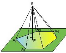
Detachment Sailboats (Velella). The representative is a sea boat. This is a large floating polyp, facing down with tentacles. A triangular hollow sail (Fig. 88) is formed from its chitinoid hydrotheque, holding the polyp like a float at the surface of the water. On the lower surface of the polyp, gonophores or jellyfish bud off.

Rice. 87. Life cycle of the freshwater hydroid jellyfish Craspedocusta (according to Naumov): 1 - egg, 2 - frustula larva, 3 - tentacleless polyps, 4 - tentacled polyps, 5 - jellyfish budding

Hydra Squad (Hydrida)- solitary freshwater polyps that develop without alternation of generations. Representative - freshwater hydra (Hydra vulgaris).
This order includes exclusively freshwater polyp species. Hydras are solitary, primitive polyps. There are few of them (15-20 species), but they are widely distributed throughout the world. Freshwater hydras are small polyps (on average, from a few millimeters to 3 cm in length) that attach themselves to freshwater plants. They are usually found attached to the underside of submerged or floating leaves. The first sketches of the hydra were made by the inventor of the microscope A. Leeuwenhoek in the 17th century. But these animals became widely known only after the publication of the works of the Swiss teacher and naturalist R. Tremblay (1710-1784). He discovered the green hydra, later named Chlorohydra. P. Tremblay published a book on the structure and life of the hydra, in which he proved its animal nature. He made observations on the nutrition of hydras, which actively captured small crustaceans with tentacles. Another merit of R. Tremblay was the conduct of classical experiments on the regeneration of hydra. For the first time it was proved that such low-organized multicellular organisms,
like hydra, they are able to regenerate even from small cut off parts of the body. For the ability to restore the cut off front ("head") section of the body, these animals were named by K. Linnaeus hydras (Hydra) in honor of the mythical creature - the many-headed hydra, capable of regrowing lost heads.
Development is direct, without the formation of larvae.
Subclass Siphonophora (Siphonophora)
Siphonophores are polymorphic colonial hydroids. Siphonophores differ from polymorphic marine hydroid polyps (Leptolida) in that their diversity of individuals in the colony is associated with functional differentiation not only of polypoid individuals, but also of medusoid ones. Siphonophores are exclusively marine floating colonial hydroids. They are varied in shape and size. The largest of them reach 2-3 m in length, and the smallest - about 1 cm.
Structure and functions. Each siphonophore colony consists of a trunk, on which individual individuals are located, performing different functions (Fig. 89). The trunk of the colony is hollow and connects the gastric cavities of all individuals into one gastrovascular system. At the top of the colony is an air bladder pneumatophore. This is a modified medusoid specimen that performs the function of a float, sail and hydrostatic apparatus. Special gas cells inside the pneumatophore are able to release gas that fills its gastric cavity. The composition of the gas inside the pneumatophore is close to air, but it has a higher nitrogen content, carbon dioxide and below - oxygen. When the pneumatophore is filled with gas, the colony stays close to the surface of the water. During a storm, the walls of the pneumatophore contract, and the gas is released through the pore to the outside. In this case, the pneumatophore decreases, the specific gravity of the colony increases, and it sinks into the depth. Under the pneumatophore is a group of swimming bells - nektophores. These medusoids lack an oral stalk, tentacles, and sensory organs. Their function is motor. By shortening the sail, the umbrellas of some nektophores either fill with water or throw out portions of water, which ensures the “reactive” movement of the colony forward by the pneumatophore.
On the rest of the trunk there are complexes of individuals with different functions - cormidia. The composition of the cormidium may include the following individuals: operculum, gastrozoid, palpon, cystozoid, gonophore. Lid- a modified flattened polyp covering the cormidia. Gastrozoid is a lactating polyp with a mouth. He is accompanied by a polyp, modified into a noose, seated with stinging cells. The food captured by the gastrozoids is then distributed through the gastro-vascular system among all members of the colony. Palpons present

Rice. 89. Scheme of the structure of the siphonophore (according to Kholodkovsky): 1 - pneumatophore, 2 - nektophore, 3 - gonophore, 4 - gastrozoid, 5 - noose, 6 - operculum, 7 - palpon, 8 - colony trunk

Rice. 90. Siphonophores: A - Portuguese boat Physalia physalis, B - Physophora hydrostatica (according to Kestner)
a modified polyp without a mouth opening. Recently, it turned out that they perform the function of intracellular digestion. From the cavity of the colony stem, food particles enter the palpons, where they are assimilated by endoderm cells. Another derivative of polyps are cystozoids with an excretory pore instead of a mouth. These are individuals with an excretory function. Finally, sexual individuals are permanent members of Cormidium - gonophores. These are modified medusoids with sexual products. Colonies may be heterosexual or bisexual. In some siphonophores, jellyfish bud and then an alternation of generations of a "polymorphic colony and jellyfish appears. Fertilization is external. Sex cells enter the water. Planulas develop from fertilized eggs, which are first converted into a single individual, and then into a colony.
A spectacular representative of the siphonophore is the Portuguese boat - physalia (Physaha, Fig. 90). This is a large view from the warm seas
with a pneumatophore up to 30 cm and long tentacles up to 2-3 m. Physalia is a poisonous coelenterate. The stinging cells of the physalia paralyze even such large prey as fish. Burns from physalia are also dangerous for humans. The pneumatophores of the physalia are pink or blue. They are thin, but very strong, as they consist of two layers of ectoderm, endoderm and mesoglea as a result of the formation of a double wall, and are also covered on top with a chitinoid membrane secreted by the ectoderm. On the pneumatophore there is a comb having a curved S-shape. This is a kind of sail of the colony. Under the influence of the wind, "Portuguese boats" drift on the surface of the sea.
Origin of siphonophore. Such complex polymorphic colonies as siphonophores, in which individual individuals are similar to organs in other multicellular organisms, are considered by some scientists to be a single organism. However, most researchers consider the siphonophore as a complex and perfect colony of multicellular organisms. Proof of this is the smooth transition in the hydroid class from single to colonial polyps, from monomorphic to dimorphic and polymorphic colonies. Forms similar to the siphonophore already exist in the subclass of hydroids (Velella). Here the evolutionary phenomena of polymerization and oligomerization according to V. A. Dogel (1882-1955) take place. The evolutionary transition of hydroids to coloniality with the formation of many individuals in a colony is a manifestation of the principle of polymerization. And the functional specialization of individuals in the colony with a decrease in the number of functions, complication of the structure, and an increase in the integration of individuals is the result of the oligomerization process.
Coelenterates are the first two-layered ancient animals with radial symmetry, an intestinal (gastric) cavity and a mouth opening. They live in water. There are sessile forms (benthos) and floating (plankton), which is especially pronounced in jellyfish. Predators feeding on small crustaceans, fish fry, aquatic insects.
A significant role in the biology of the southern seas is played by coral polyps, which form reefs and atolls, which serve as shelters and spawning grounds for fish; at the same time they pose a danger to ships.
Large jellyfish are eaten by people, but they also cause serious burns to swimmers. Reef limestone is used for decoration and as a building material. However, destroying reefs, a person reduces fish wealth. The most famous reefs in south seas- along the coast of Australia, off the Sunda Islands, in Polynesia.
Intestinal - the oldest type of primitive two-layer multicellular animals. Lacking true organs. Their study is of exceptional importance for understanding the evolution of the animal world: the ancient species of this type were the progenitors of all higher multicellular animals.
Intestinal - predominantly marine, less often freshwater animals. Many of them are attached to underwater objects, others slowly swim in the water. Attached forms are usually goblet-shaped and are called polyps. They are attached to the substrate with the lower end of the body, at the opposite end there is a mouth surrounded by a corolla of tentacles. The floating forms are usually bell-shaped or umbrella-shaped and are called jellyfish.
The body of the coelenterates has radial (radial) symmetry. Two or more (2, 4, 6, 8 or more) planes can be drawn through it, dividing the body into symmetrical halves. In the body, which can be compared with a two-layer bag, only one cavity is developed - the gastric cavity, which acts as a primitive intestine (hence the name of the type). It communicates with the external environment through a single opening that functions as a mouth and anus. The sac wall consists of two cell layers: the outer or ectoderm and the inner or endoderm. A structureless substance lies between the cell layers. It forms either a thin supporting plate or a wide layer of gelatinous mesoglea. In many coelenterates (for example, jellyfish), channels depart from the gastric cavity, which together with the gastric cavity form a complex gastrovascular (gastrointestinal) system.

The cells of the body of the coelenterates are differentiated.
- Ectoderm cells
represented by several types:
- integumentary (epithelial) cells - form the cover of the body, perform a protective function
Epithelial muscle cells - in the lower forms (hydroid) integumentary cells have a long process extended parallel to the surface of the body, in the cytoplasm of which contractile fibrils are developed. The totality of such processes forms a layer of muscular formations. Epithelial muscle cells combine the functions of a protective cover and a motor apparatus. Due to the contraction or relaxation of muscle formations, the hydra can shrink, thicken or narrow, stretch, bend to the side, attach to other parts of the stems and thus move slowly. In higher intestinal cavities, muscle tissue is isolated. Jellyfish have powerful bundles of muscle fibers.
- stellate nerve cells. The processes of nerve cells communicate with each other, forming the nerve plexus, or diffuse nervous system.
- intermediate (interstitial) cells - restore damaged areas of the body. Intermediate cells can form integumentary-muscular, nervous, sex and other cells.
- stinging (nettle) cells - located among the integumentary cells singly or in groups. They have a special capsule in which lies a spirally twisted stinging thread. The cavity of the capsule is filled with liquid. On the outer surface of the stinging cell, a thin sensitive hair is developed - the cnidocil. When touched by a small animal, the hair is deflected, and the stinging thread is thrown out and straightens, through which a paralyzing poison enters the body of the prey. After the thread is ejected, the stinging cell dies. Stinging cells are renewed due to undifferentiated interstitial cells lying in the ectoderm.
- integumentary (epithelial) cells - form the cover of the body, perform a protective function
- Endoderm cells
line the gastric (intestinal) cavity and perform mainly the function of digestion. These include
- glandular cells that secrete digestive enzymes into the gastric cavity
- digestive cells with phagocytic function. Digestive cells (in lower forms) also have processes in which contractile fibers are developed, oriented perpendicular to similar formations of integumentary muscle cells. Flagella are directed from the epithelial-muscular cells towards the intestinal cavity (1-3 from each cell) and outgrowths can form, resembling false legs, which capture small food particles and digest them intracellularly in digestive vacuoles. Thus, in intestinal cavities, intracellular digestion characteristic of protozoa is combined with intestinal digestion characteristic of higher animals.
The nervous system is primitive. In both cell layers there are special sensitive (receptor) cells that perceive external stimuli. A long nerve process departs from their basal end, along which the nerve impulse reaches multi-processed (multipolar) nerve cells. The latter are located one by one, do not form nerve nodes, but are connected with each other by their processes and make up the nervous network. Such a nervous system is called diffuse.
The reproductive organs are represented only by the sex glands (gonads). Reproduction occurs sexually and asexually (budding). For many coelenterates, alternation of generations is characteristic: polyps, multiplying by budding, give both new polyps and jellyfish. The latter, reproducing sexually, give a generation of polyps. This alternation of sexual reproduction with vegetative reproduction is called metagenesis. [show] .
Metagenesis occurs in many coelenterates. For example, the well-known Black Sea jellyfish - Aurelia - reproduces sexually. The spermatozoa and eggs that arise in her body are released into the water. From fertilized eggs, individuals of the asexual generation develop - Aurelia polyps. The polyp grows, its body lengthens, and then is divided by transverse constrictions (polyp strobilation) into a number of individuals that look like saucers stacked in a stack. These individuals separate from the polyp and turn into sexually reproducing jellyfish.
Systematically, the type is divided into two subtypes: cnidarians (Cnidaria) and non-cnidators (Acnidaria). There are about 9,000 known species of cnidarians, and only 84 species of non-cnidaria.
SUBTYPE CIDING
Subtype characteristic
Intestinal, called stingers, have stinging cells. These include the classes: hydroid (Hydrozoa), scyphoid (Scyphozoa) and coral polyps (Anthozoa).
Class hydroid (Hydrozoa)
A single individual has the form of either a polyp or a jellyfish. The intestinal cavity of polyps is devoid of radial partitions. Sex glands develop in the ectoderm. About 2800 species live in the sea, but there are several freshwater forms.
- Subclass Hydroids (Hydroidea) - bottom colonies, adherent. In some non-colonial species, polyps are able to swim near the surface of the water. Within each species, all individuals of the medusoid structure are the same.
- Order Leptolida (Leptolida) - there are individuals of both polypoid and medusoid origin. Mostly marine, very rarely freshwater organisms.
- Hydrocoral detachment (Hydrocorallia) - the trunk and branches of the colony are calcareous, often painted in a beautiful yellowish, pink or red color. Medusoid individuals are underdeveloped and immersed deep in the skeleton. Exclusively marine organisms.
- Detachment Chondrophora (Chondrophora) - the colony consists of a floating polyp and medusoid individuals attached to it. Exclusively marine animals. Previously classified as a subclass of siphonophores.
- Detachment Tachilida (Trachylida) - exclusively marine hydroid, shaped like a jellyfish, no polyps.
- Order Hydra (Hydrida) - single freshwater polyps, do not form jellyfish.
- Subclass Siphonophora (Siphonophora) - floating colonies, which include polypoid and medusoid individuals of various structures. They live exclusively in the sea.
Hydra freshwater polyp- a typical representative of hydroids, and at the same time all cnidaria. Several species of these polyps are widely distributed in ponds, lakes and small rivers.
Hydra is a small, about 1 cm long, brownish-green animal with a cylindrical body. At one end there is a mouth surrounded by a corolla of very mobile tentacles, which vary from 6 to 12 in different species. At the opposite end there is a stem with a sole that serves to attach to underwater objects. The pole on which the mouth is located is called oral, the opposite is called aboral.
Hydra leads a sedentary lifestyle. Attached to underwater plants and hanging into the water with its mouth end, it paralyzes passing prey with stinging threads, captures it with tentacles and sucks it into the gastric cavity, where digestion occurs under the action of glandular cell enzymes. Hydras feed mainly on small crustaceans (daphnia, cyclops), as well as ciliates, oligochaete worms and fish fry.
Digestion. Under the action of enzymes of the glandular cells of the endoderm lining the gastric cavity, the body of the captured prey breaks up into small particles that are captured by cells that have pseudopodia. Some of these cells are in their permanent place in the endoderm, others (amoeboid) are mobile and move. These cells complete the digestion of food. Consequently, there are two ways of digestion in coelenterates: along with the more ancient, intracellular, an extracellular, more progressive way of processing food appears. Subsequently, in connection with the evolution of the organic world and the digestive system, intracellular digestion lost its significance in the act of nutrition and assimilation of food, but the ability for it was preserved in individual cells in animals at all stages of development up to the highest, and in humans. These cells, discovered by I. I. Mechnikov, were called phagocytes.
Due to the fact that the gastric cavity ends blindly and the anus is absent, the mouth serves not only for eating, but also for removing undigested food residues. The gastric cavity performs the function of blood vessels (moving nutrients through the body). The distribution of substances in it is ensured by the movement of flagella, which are provided with many endodermal cells. The contractions of the whole body serve the same purpose.

Respiration and excretion carried out by diffusion by both ectodermal and endodermal cells.
Nervous system. Nerve cells form a network throughout the hydra's body. This network is called the primary diffuse nervous system. There are especially many nerve cells around the mouth, on the tentacles and soles. Thus, the simplest coordination of functions appears in the coelenterates.
sense organs. Not developed. Touch with the entire surface, the tentacles (sensitive hairs) are especially sensitive, throwing out stinging threads that kill prey.
Hydra locomotion carried out by transverse and longitudinal muscle fibers included in epithelial cells.
Hydra Regeneration- restoration of the integrity of the hydra body after its damage or loss of part of it. A damaged hydra regenerates lost body parts not only after it has been cut in half, but even if it has been divided into a huge number of parts. A new animal is able to grow from 1/200 of a hydra, in fact, a whole organism is restored from a grain. Therefore, hydra regeneration is often called an additional method of reproduction.
reproduction. Hydra reproduces asexually and sexually.
During the summer, hydra reproduces asexually - by budding. In the middle part of her body is a budding belt, on which tubercles (buds) form. The kidney grows, a mouth and a tentacle are formed on its top, after which the kidney is laced at the base, separated from the mother's body and begins to live on its own.
With the approach of cold weather in autumn, germ cells - eggs and spermatozoa - are formed from intermediate cells in the hydra ectoderm. The eggs are closer to the base of the hydra, the spermatozoa develop in the tubercles (male gonads) located closer to the mouth. Each spermatozoon has a long flagellum, with the help of which it swims in the water, reaches the egg and fertilizes it in the mother's body. The fertilized egg begins to divide, becomes covered with a dense double shell, sinks to the bottom of the reservoir and overwinter there. In late autumn, adult hydras die. In the spring, a new generation develops from overwintered eggs.
Colonial polyps(for example, the colonial hydroid polyp Obelia geniculata) live in the seas. A single individual of the colony, or the so-called hydrant, is similar in structure to the hydra. The wall of its body, like the hydra, consists of two layers: endoderm and ectoderm separated by a jelly-like structureless mass called mesoglea. The body of the colony is a branched coenosarc, inside of which there are separate polyps, interconnected by outgrowths of the intestinal cavity into a single digestive system, which makes it possible to distribute the food captured by one polyp among the members of the colony. Outside, the coenosarc is covered with a hard shell - the perisarc. Near each hydrant, this shell forms an extension in the form of a goblet - a hydrotech. The corolla of tentacles can be drawn into the extension when stimulated. The mouth opening of each hydrant is located on an outgrowth, around which there is a corolla of tentacles.
Colonial polyps reproduce asexually by budding. At the same time, the individuals that developed on the polyp do not come off, like in the hydra, but remain associated with the maternal organism. An adult colony looks like a bush and consists mainly of two types of polyps: gastrozoids (hydrants), which provide food and protect the colony with stinging cells on tentacles, and gonozoids, which are responsible for reproduction. There are also polyps specialized to perform a protective function.
A gonozoid is a rod-shaped formation elongated in length with an extension at the top, without a mouth opening and tentacles. Such an individual cannot feed on its own; it receives food from hydrants through the gastric system of the colony. This formation is called a blastostyle. The skeletal membrane gives a bottle-shaped extension around the blastostyle - the gonotheca. All this formation as a whole is called gonangia. In the gonangium, on the blastostyle, jellyfish are formed by budding. They bud from the blastostyle, emerge from the gonangium, and begin to lead a free lifestyle. As the jellyfish grows, germ cells form in its gonads, which are released into the external environment, where fertilization occurs.
A blastula is formed from a fertilized egg (zygote), with the further development of which a two-layer larva, planula, freely floating in water, covered with cilia, is formed. The planula settles to the bottom, attaches to underwater objects and continues to grow and gives rise to a new polyp. This polyp forms a new colony by budding.

Hydroid jellyfish have the shape of a bell or an umbrella, from the middle of the ventral surface of which hangs a trunk (oral stalk) with a mouth opening at the end. Along the edge of the umbrella are tentacles with stinging cells and sticky pads (suckers) that serve to catch prey (small crustaceans, larvae of invertebrates and fish). The number of tentacles is a multiple of four. Food from the mouth enters the stomach, from which four straight radial canals extend, encircling the edge of the jellyfish umbrella (the annular canal of the intestine). The mesoglea is much better developed than that of the polyp, and makes up the bulk of the body. This is due to the greater transparency of the body. The way the jellyfish moves is "reactive", this is facilitated by a fold of ectoderm along the edge of the umbrella, called the "sail".
In connection with the free way of life, the nervous system of jellyfish is better developed than that of polyps, and, in addition to the diffuse nervous network, it has accumulations of nerve cells along the edge of the umbrella in the form of a ring: external - sensitive and internal - motor. The sensory organs are also located here, represented by light-sensitive eyes and statocysts (organs of balance). Each statocyst consists of a vesicle with a calcareous body - statolith, located on elastic fibers coming from the sensitive cells of the vesicle. If the position of the jellyfish's body in space changes, the statolith shifts, which is perceived by sensitive cells.
Jellyfish have separate sexes. Their gonads are located under the ectoderm, on the concave surface of the body under the radial canals, or in the region of the oral proboscis. Sex cells are formed in the gonads, which, when mature, are excreted through a gap in the body wall. biological significance mobile jellyfish is that thanks to them, the settlement of hydroids occurs.
Scyphozoa class
An individual has the appearance of either a small polyp or a large jellyfish, or the animal bears signs of both generations. The intestinal cavity of polyps has 4 incomplete radial septa. Sex glands develop in the endoderm of jellyfish. About 200 species. Exclusively marine organisms.
- Order Coronomedusa (Coronata) - mainly deep-sea jellyfish, the umbrella of which is divided by a constriction into a central disk and a crown. The polyp forms a protective chitinoid tube around itself.
- Detachment Discomedusae (Discomedusae) - the umbrella of jellyfish is solid, there are radial channels. Polyps lack a protective tube.
- Detachment of Cubomedusae (Cubomedusae) - the jellyfish umbrella is solid, but devoid of radial channels, the function of which is performed by far protruding pockets of the stomach. Polyp without protective tube.
- The detachment of Stauromedusae (Stauromedusae) is a kind of benthic organisms that combine in its structure the signs of a jellyfish and a polyp.
Most of the life cycle of coelenterates from this class takes place in the medusoid phase, while the polypoid phase is short-lived or absent. Scyphoid coelenterates have a more complex structure than hydroid ones.

Unlike hydroid jellyfish, scyphoid jellyfish are larger, have a highly developed mesoglea, and a more developed nervous system with clusters of nerve cells in the form of nodules - ganglia, which are located mainly around the circumference of the bell. The stomach cavity is divided into chambers. Canals extend radially from it, united by an annular canal located along the edge of the body. The collection of channels forms the gastrovascular system.
The mode of movement is "reactive", but since the Scyphoid do not have a "sail", movement is achieved by shortening the walls of the umbrella. Along the edge of the umbrella are complex sense organs - ropalia. Each ropalium contains an "olfactory fossa", an organ of balance and stimulation of the movement of the umbrella - statocysts, a light-sensitive eye. Scyphoid jellyfish are predators, but deep-sea species feed on dead organisms.
Sex cells are formed in the sex glands - gonads located in the endoderm. Gametes are shed through the mouth, and planula develops from fertilized eggs. Further development proceeds with alternation of generations, and the generation of jellyfish prevails. The generation of polyps is short-lived.
The tentacles of jellyfish are equipped with a large number of stinging cells. The burns of many jellyfish are sensitive to large animals and humans. Severe burns with severe consequences can be caused by a polar jellyfish of the genus Cyanea, reaching a diameter of 4 m, with tentacles up to 30 m long. Bathers in the Black Sea are sometimes burned by the jellyfish Pilema pulmo, and in the Sea of Japan - gonionemus (Gonionemus vertens).
Representatives of the class of scyphoid jellyfish include:
- aurelia jellyfish (eared jellyfish) (Aurelia aurita) [show]
.
Aurelia long-eared jellyfish (Aurelia aurita)
It lives in the Baltic, White, Barents, Black, Azov, Japanese and Bering, and is often found in mass quantities.
It got its name due to the oral lobes, resembling donkey ears in shape. The umbrella of an eared jellyfish sometimes reaches 40 cm in diameter. It is easily recognizable by its pinkish or slightly purple color and four dark horseshoes in the middle part of the umbrella - the gonads.
In summer, in calm calm weather, at low tide or high tide, you can see a large number of these beautiful jellyfish, slowly carried by the current. Their bodies gently sway in the water. The eared jellyfish is a poor swimmer, thanks to the contractions of the umbrella, it can only slowly rise to the surface, and then, frozen motionless, sink into the depths.
On the edge of the aurelia umbrella there are 8 ropalia bearing eyes and statocysts. These sense organs allow the jellyfish to keep a certain distance from the surface of the sea, where its delicate body will quickly be torn apart by waves. The eared jellyfish captures food with the help of long and very thin tentacles, which "sweep" small planktonic animals into the jellyfish's mouth. Swallowed food first enters the throat, and then into the stomach. From here, 8 straight radial canals and the same number of branching canals originate. If, using a pipette, a carcass solution is introduced into the stomach of a jellyfish, then one can trace how the flagellar epithelium of the endoderm drives food particles through the channels of the gastric system. First, the ink penetrates into the non-branching canals, then it enters the annular canal and returns back to the stomach through the branching canals. From here, undigested food particles are thrown out through the mouth opening.
The sex glands of Aurelia, having the shape of four open or complete rings, are located in the pockets of the stomach. When the eggs in them mature, the wall of the gonad ruptures and the eggs are thrown out through the mouth. Unlike most scyphomedusae, Aurelia shows a kind of concern for offspring. The oral lobes of this jellyfish carry a deep longitudinal groove along their inner side, starting from the mouth opening and passing to the very end of the blade. On both sides of the gutter are numerous small holes that lead to small cavities-pockets. In a floating jellyfish, its oral lobes are lowered, so that the eggs emerging from the mouth opening inevitably fall into the gutters and, moving along them, linger in pockets. This is where the eggs are fertilized and developed. From pockets, fully formed planulae come out. If you place a large female Aurelia in an aquarium, then after a few minutes you can notice a lot of bright dots in the water. These are planulas that have left their pockets and float with the help of cilia.
Young planulas show an urge to move towards the light source, soon they accumulate in the upper part of the illuminated side of the aquarium. Probably, this property helps them to get out of the darkened pockets into the wild and stay close to the surface without going into the depths.
Soon, the planula tends to sink to the bottom, but always in bright places. Here they continue to swim briskly. The period of free-moving life of the planula lasts from 2 to 7 days, after which they settle to the bottom and attach their front end to some solid object.
After two or three days, the settled planula turns into a small polyp - a scyphist, which has 4 tentacles. Soon, 4 new tentacles appear between the first tentacles, and then 8 more tentacles. Scyphistomas actively feed, capturing ciliates and crustaceans. Cannibalism is also observed - eating planulas of the same species by scyphistomas. Scyphistomas can reproduce by budding, forming similar polyps. The scyphistoma hibernates, and next spring, with the onset of warming, serious changes occur in it. The tentacles of the scyphistoma shorten, and ring-shaped constrictions appear on the body. Soon, the first ether separates from the upper end of the scyphistoma - a small, completely transparent, star-shaped jellyfish larva. By the middle of summer, a new generation of eared jellyfish develops from the ether.
- jellyfish cyanea (Suapea) [show]
.

Scyphoid jellyfish cyanide - is the largest jellyfish. These giants among the intestinal cavities live only in cold waters. The diameter of the cyanide umbrella can reach 2 m, the length of the tentacles is 30 m. Outwardly, the cyanide is very beautiful. The umbel is usually yellowish in the center, dark red towards the edges. The mouth lobes look like wide crimson-red curtains, the tentacles are painted in a light pink color. Young jellyfish are especially bright in color. The poison of stinging capsules is dangerous for humans.
- jellyfish rhizostoma, or cornerot (Rhizostoma pulmo) [show]
.

Scyphoid jellyfish cornerot lives in the Black and Azov Seas. The umbrella of this jellyfish is hemispherical or conical in shape with a rounded top. Large specimens of rhizostomy are difficult to fit in a bucket. The color of the jellyfish is whitish, but a very bright blue or purple border runs along the edge of the umbrella. This jellyfish has no tentacles, but its oral lobes branch in two, and their lateral sides form numerous folds and grow together. The ends of the oral lobes do not bear folds and end with eight root-like outgrowths, from which the jellyfish got its name. The mouth of adult Cornerots is overgrown, and its role is played by numerous small holes in the folds of the oral lobes. Here, in the oral lobes, digestion also occurs. In the upper part of the oral lobes of the cornerot there are additional folds, the so-called epaulettes, which enhance the digestive function. Cornerots feed on the smallest planktonic organisms, sucking them together with water into the gastric cavity.
Cornerots are pretty good swimmers. The streamlined shape of the body and the strong musculature of the umbrella allow them to move forward with quick, frequent jerks. It is interesting to note that, unlike most jellyfish, Cornerot can change its movement in any direction, including down. Bathers are not very happy with the meeting with the cornerot: touching it, you can get a rather strong painful "burn". Cornerots usually keep at a shallow depth near the coast, often found in large numbers in the Black Sea estuaries.
- edible ropilema (Rhopilema esculenta) [show]
.
Edible Ropilema (Rhopilema esculenta) lives in warm coastal waters, accumulating in masses near river mouths. It has been observed that these jellyfish grow most intensively after the onset of the summer tropical rainy season. During the rainy season, rivers carry a large amount of organic matter into the sea, contributing to the development of plankton, which jellyfish feed on. Along with Aurelia, ropilema is eaten in China and Japan. Outwardly, ropilema resembles the Black Sea cornerot, differs from it in the yellowish or reddish color of the oral lobes and the presence of a large number of finger-like outgrowths. The mesoglea of the umbrella is used for food.
Ropilemas are immobile. Their movements depend mainly on sea currents and winds. Sometimes, under the influence of current and wind, clusters of jellyfish form belts 2.5-3 km long. In some parts of the coast of South China, the sea turns white in summer from the accumulated ropils, which sway near the surface.
They catch jellyfish with nets or special fishing gear, which look like a large bag of fine-mesh net, worn on a hoop. During high or low tide, the bag is inflated by the current and jellyfish enter it, which cannot get out due to their inactivity. In the extracted jellyfish, the oral lobes are separated and the umbrella is washed until the internal organs and mucus are completely removed. Thus, in fact, only the mesoglea of the umbrella enters further processing. By figurative expression Chinese, the meat of jellyfish is "crystal". Jellyfish are salted with table salt mixed with alum. Salted jellyfish are added to various salads, and are also eaten boiled and fried, seasoned with pepper, cinnamon and nutmeg. Of course, a jellyfish is a low-nutrient product, but nevertheless, salted ropilemas contain a certain amount of proteins, fats and carbohydrates, as well as vitamins B 12, B 2 and nicotinic acid.
The eared jellyfish, the edible ropilema, and some species of scyphomedusa close to it are, in all likelihood, the only coelenterates that are eaten by humans. In Japan and China there is even a special fishery for these jellyfish, and thousands of tons of "crystal meat" are mined there annually.
Class coral polyps (Anthozoa)
Coral polyps are exclusively marine organisms of a colonial or sometimes solitary form. About 6,000 species are known. In size, coral polyps are larger than hydroids. The body has a cylindrical shape and is not divided into a trunk and a leg. In colonial forms, the lower end of the polyp body is attached to the colony, while in single polyps it is provided with an attachment sole. The tentacles of coral polyps are located in one or more closely spaced corollas.
There are two large groups coral polyps: eight-rayed (Octocorallia) and six-rayed (Nehasorallia). The former always have 8 tentacles, and they are equipped with small outgrowths at the edges - pinnules, in the latter the number of tentacles is usually quite large and, as a rule, a multiple of six. The tentacles of six-pointed corals are smooth, without pinnules.
The upper part of the polyp, between the tentacles, is called the oral disc. In its middle is a slit-like mouth opening. The mouth leads to the pharynx lined with ectoderm. One of the edges of the oral fissure and the pharynx descending from it is called the siphonoglyph. The ectoderm of the siphonoglyph is covered with epithelial cells with very large cilia, which are in constant motion and drive water into the intestinal cavity of the polyp.
The intestinal cavity of the coral polyp is divided into chambers by longitudinal endodermal septa (septa). In the upper body of the polyp, the septa grow with one edge to the body wall, and with the other to the pharynx. In the lower part of the polyp, below the pharynx, the septa are attached only to the body wall, as a result of which the central part of the gastric cavity - the stomach - remains undivided. The number of septa corresponds to the number of tentacles. On each septum, along one of its sides, there is a muscular roller.
The free edges of the septa are thickened and are called mesenteric filaments. Two of these filaments, located on a pair of adjacent septa that oppose the siphonoglyph, are covered with special cells bearing long cilia. The cilia are in constant motion and drive water out of the gastric cavity. The joint work of the ciliary epithelium of these two mesenteric filaments and the siphonoglyph ensures a constant change of water in the gastric cavity. Thanks to them, fresh, oxygen-rich water constantly enters the intestinal cavity. Species that feed on the smallest planktonic organisms also receive food. The remaining mesenteric filaments play important role in digestion, as they are formed by glandular endodermal cells that secrete digestive juices.
Reproduction is asexual - by budding, and sexual - with metamorphosis, through the stage of a free-swimming larva - planula. The sex glands develop in the endoderm of the septum. For coral polyps, only the polypoid state is characteristic, there is no alternation of generations, since they do not form jellyfish and, accordingly, the medusoid stage is absent.
The ectoderm cells of coral polyps produce horny matter or secrete carbonic lime, from which the external or internal skeleton is built. In coral polyps, the skeleton plays a very important role.
Eight-ray corals have a skeleton consisting of individual calcareous needles - spicules located in the mesoglea. Sometimes spicules are interconnected, merging or uniting with an organic horn-like substance.
Among six-pointed corals there are non-skeletal forms, such as sea anemones. More often, however, they have a skeleton, and it can be either internal - in the form of a rod of a horn-like substance, or external - calcareous.
The skeleton of representatives of the Madreporaceae group reaches especially great complexity. It is secreted by the ectoderm of polyps and at first looks like a plate or a low cup in which the polyp itself sits. Further, the skeleton begins to grow, radial ribs appear on it, corresponding to the septa of the polyp. Soon the polyp turns out to be, as it were, impaled on a skeletal base, which protrudes deeply from below into its body, although it is delimited everywhere by the ectoderm. The skeleton of stony corals is very strongly developed: soft tissues cover it in the form of a thin film.
The skeleton of the coelenterates plays the role of a support system, and together with the stinging apparatus it represents a powerful defense against enemies, which contributed to their existence over long geological periods.
- Subclass Eight-beam corals (Octocorallia) - colonial forms, as a rule, adherent to the ground. The polyp has 8 tentacles, eight septa in the gastric cavity, and an internal skeleton. On the sides of the tentacles there are outgrowths - pinnules. This subclass is subdivided into units:
- Order Solar corals (Helioporida) - solid, massive skeleton.
- Order Alcyonaria - soft corals, skeleton in the form of calcareous needles [show]
.
Most alcyonaria are soft corals that do not have a pronounced skeleton. Only some tubipores have a developed calcareous skeleton. In the mesoglea of these corals, tubules are formed, which are soldered to each other by transverse plates. The skeleton vaguely resembles an organ in shape, so tubipores have another name - organ. Organs are involved in the process of reef formation.
- Order Horn corals (Gorgonaria) - a skeleton in the form of calcareous needles, usually there is also an axial skeleton of horn-like or calcified organic matter passing through the trunk and branches of the colony. This order includes red, or noble coral (Corallium rubrum), which is the object of fishing. Jewelry is made from the skeletons of red coral.
- Order Sea feathers (Rennaturia) - a kind of colony consisting of a large polyp, on the lateral outgrowths of which secondary polyps develop. The base of the colony is embedded in the ground. Some species are able to move.
- Subclass Six-beam corals (Hexacorallia) - colonial and solitary forms. Tentacles without lateral outgrowths, their number is usually equal to or a multiple of six. The gastric cavity is divided complex system partitions, the number of which is also a multiple of six. Most of the representatives have an external calcareous skeleton, there are groups that are devoid of a skeleton. Includes:

SUBTYPE CLEAR
Subtype characteristic
Non-stinging coelenterates instead of stinging ones have special sticky cells on their tentacles that serve to capture prey. This subtype includes a single class - ctenophores.
Ctenophore class (Ctenophora)- unites 90 species of marine animals with a translucent sac-like gelatinous body, in which the channels of the gastrovascular system branch. Along the body there are 8 rows of rowing plates, consisting of fused large cilia of ectoderm cells. There are no stinging cells. On the sides of the mouth there is one tentacle each, due to which a two-beam type of symmetry is created. Ctenophores always swim forward with the oral pole, using rowing plates as an organ of propulsion. The mouth opening leads to the ectodermal pharynx, which passes into the esophagus. Behind it is the endodermal stomach with radial canals extending from it. At the aboral pole there is a special organ of balance called the aboral. It is built on the same principle as jellyfish statocysts.
Ctenophores are hermaphrodites. The sex glands are located on the processes of the stomach under the rowing plates. The gametes are brought out through the mouth. In the larvae of these animals, the formation of the third germ layer, the mesoderm, can be traced. This is an important progressive feature of ctenophores.
Ctenophores are of great interest from the point of view of the phylogenesis of the animal world, because in addition to the most important progressive feature - the development between the ecto- and endoderm of the rudiment of the third germ layer - the mesoderm, due to which numerous muscle elements develop in the gelatinous substance of the mesoglea in adult forms, they have a number of other progressive features , bringing them closer to higher types of multicellular organisms.
The second progressive feature is the presence of elements of bilateral (bilateral) symmetry. It is especially clear in the crawling comb jelly Coeloplana metschnikowi, studied by A.O. Kovalevsky, and Ctenoplana kowalewskyi, discovered by A.A. Korotnev (1851-1915). These ctenophores have a flattened shape and, in adulthood, lack rowing plates, and therefore can only crawl along the bottom of the reservoir. The side of the body of such a ctenophore facing the ground becomes ventral (ventral); the sole develops on it; the opposite, upper side of the body becomes the dorsal or dorsal side.
Thus, in the phylogenesis of the animal world, the ventral and dorsal sides of the body became distinct for the first time in connection with the transition from swimming to crawling. There is no doubt that modern crawling ctenophores have retained in their structure the progressive features of that group of ancient coelenterates that became the ancestors of higher types of animals.
However, in his detailed studies, V.N. Beklemishev (1890-1962) showed that despite the common features of the structure of ctenophores and some marine flatworms, the assumption of the origin of flatworms from ctenophores is untenable. The common features of their structure are due to the general conditions of existence, which lead to a purely external, convergent similarity.
The value of coelenterates
Colonies of hydroids, attached to various underwater objects, often grow very densely on the underwater parts of ships, covering them with a shaggy "fur coat". In these cases, hydroids bring significant harm to navigation, since such a "fur coat" sharply reduces the speed of the vessel. There are many cases when hydroids, settling inside the pipes of the sea water supply, almost completely closed their gap and prevented the supply of water. It is rather difficult to fight hydroids, since these animals are unpretentious and develop quite well, it would seem, in adverse conditions. In addition, they are characterized by rapid growth - bushes 5-7 cm tall grow in a month. To clear the bottom of the ship from them, you have to put it in a dry dock. Here the ship is cleared of overgrown hydroids, polychaetes, bryozoans, sea acorns and other fouling animals. Recently, special poisonous paints have been used - the underwater parts of the ship covered with them are subject to fouling to a much lesser extent.
In the thickets of hydroids living at great depths, live worms, mollusks, crustaceans, echinoderms. Many of them, such as sea goat crustaceans, find refuge among hydroids, others, such as sea "spiders" (multi-legged), not only hide in their thickets, but also feed on hydropolyps. If you move around the settlements of hydroids with a small-meshed net or, even better, use a special, so-called plankton net for this, then among the mass of small crustaceans and larvae of various other invertebrates, hydroid jellyfish will come across. Despite their small size, hydroid jellyfish are very voracious. They eat a lot of crustaceans and therefore are considered harmful animals - competitors of plankton-eating fish. Plentiful food is necessary for jellyfish for the development of reproductive products. Swimming, they scatter a huge number of eggs into the sea, which subsequently give rise to the polypoid generation of hydroids.
Some jellyfish pose a serious danger to humans. Cornerot jellyfish are very numerous in the Black and Azov Seas in summer, touching them, you can get a strong and painful "burn". In the fauna of our Far Eastern seas there is also one jellyfish that causes serious diseases when it comes into contact with it. Locals call this jellyfish "cross" for the cruciform arrangement of four dark radial canals, along which four also dark-colored gonads stretch. The umbrella of the jellyfish is transparent, of a faint yellowish-green color. The size of the jellyfish is small: the umbrella of individual specimens reaches 25 mm in diameter, but usually they are much smaller, only 15-18 mm. On the edge of the umbrella of the cross (scientific name - Gonionemus vertens) there are up to 80 tentacles that can be strongly extended and contracted. The tentacles are densely seated with stinging cells, which are arranged in bands. In the middle of the length of the tentacle there is a small suction cup, with which the jellyfish is attached to various underwater objects.
Krestovichki live in the Sea of Japan and near the Kuril Islands. They usually stay in shallow water. Their favorite places are thickets of seagrass zostera. Here they swim and hang on the blades of grass, attached with their suckers. Sometimes they come across in clear water, but usually not far from the thickets of Zostera. During the rains, when the sea water off the coast is significantly desalinated, the jellyfish die. In rainy years, they are almost absent, but by the end of a dry summer, crosses appear in masses.
Although they can swim freely, they usually prefer to lie in wait for prey by attaching themselves to an object. Therefore, when one of the tentacles of the cross accidentally touches the body of a bathing person, the jellyfish rushes in this direction and tries to attach itself with the help of suction cups and stinging capsules. At this moment, the bather feels a strong "burn", after a few minutes the skin at the site of the touch of the tentacle turns red, blistered. Feeling the "burn", you must immediately get out of the water. After 10-30 minutes, general weakness sets in, back pain appears, breathing becomes difficult, arms and legs go numb. Well, if the shore is close, otherwise you can drown. The affected person should be placed comfortably and a doctor should be called immediately. For treatment, subcutaneous injections of adrenaline and ephedrine are used; in the most severe cases, artificial respiration is used. The disease lasts 4-5 days, but even after this period, people affected by a small jellyfish cannot fully recover for a long time.
Repeated burns are especially dangerous. It has been established that the poison of the cross not only does not develop immunity, but, on the contrary, makes the body hypersensitive even to small doses of the same poison. This phenomenon is known in medicine under the name of anaphylaxis.

It is quite difficult to protect yourself from the cross. In places where many people usually bathe, to combat the cross, they mow the zoster, enclose the baths with a fine-mesh net, and catch crosses with special nets.
It is interesting to note that crosses that live only in the pool have such poisonous properties. Pacific Ocean. A very close form belonging to the same species, but to a different subspecies, living on the American and European coasts Atlantic Ocean, completely harmless.
Some tropical jellyfish are eaten in Japan and China, they are called "crystal meat". The body of jellyfish has a jelly-like consistency, almost transparent, contains a lot of water and a small amount of proteins, fats, carbohydrates, vitamins B 1, B 2 and nicotinic acid.
Marine, less often freshwater animals, leading an attached lifestyle or swimming in the water. Attached forms are called polyps, floating - jellyfish.
Double layer animals, their body consists of two cell layers: the outer - ectoderm and internal - endoderm. Endoderm forms intestinal or gastric cavity. The gastric cavity communicates with environment hole that functions as oral And anal. Between the ectoderm and endoderm is mesoglea. In polyps, the mesoglea forms a base plate, while in jellyfish, it forms a thick gelatinous layer.
Ectoderm cells perform protective and motor functions. The ectoderm has special stinging cells for defense and attack. Endoderm cells line the gastric cavity and perform mainly a digestive function. Digestion intracellular And cavity.
Breathing occurs through the entire surface of the body.
Nervous system scattered or diffuse, type. Available tactile sensitivity, and in jellyfish, in connection with a floating lifestyle, light-perceiving "eyes" And balance organs.
Coelenterates have radial or radial, symmetry.
asexual reproduction budding. The sex organs are presented gonads. Fertilization is external. Some representatives are characterized by the alternation of asexual (polyp) and sexual (jellyfish) generations in the life cycle.
The type of coelenterates includes the following classes: Hydrozoa, Scyphoid jellyfish, Coral polyps.
Class Hydrozoa
freshwater hydra
A BRIEF DESCRIPTION OF
|
Habitat |
Freshwater bilayer animals. Lead an attached lifestyle |
|
Appearance |
Saccular up to 1.5 cm. Radial symmetry. The mouth at the anterior end of the body is surrounded by tentacles, the sole is the posterior end of the body, for attachment |
|
body cover |
Ectoderm - outer layer, endoderm - inner layer, mesoglea - middle layer |
|
body cavity |
There is no body cavity. There is only an intestinal cavity |
|
Digestive system |
Blindly closed intestinal cavity. The mouth opening for food intake and for the ejection of undigested food debris. Digestion intracavitary and intracellular |
|
excretorysystem |
Ectoderm cells |
|
Nervous system |
Nerve cells of stellate type. diffuse nervous system |
|
sense organs |
not developed |
|
Respiratory system |
None. Breathing through the entire surface of the body |
|
reproduction |
Asexual - by budding. Hermaphrodites. Cross fertilization. |
GENERAL CHARACTERISTICS
This class includes small forms of coelenterates. polyps And jellyfish belonging to this class are called hydroid.
Structure . The hydra's body is oblong double-layer bag, attached by the base, or sole, to the substrate (Fig. 1). outer layer - ectoderm, inner layer - endoderm. Between the layers there is a space - mesoglea.
At the free end of the body is oral cone, surrounded by a halo of 6-12 tentacles. Located on the mouth cone mouth, employee and anus. The entire surface of the body is covered ectoderm, consisting mainly of cylindrical or cuboidal epithelial cells. Their base is elongated upwards and downwards, along the longitudinal axis of the body, into a long process. The cytoplasm of the process differentiates into contractile fibers, in connection with this, the offshoot plays muscular role. Cylindrical parts of cells form single layer epithelium. Thus, cells perform a dual function - coverslip And motor and are called epithelial-muscular. With the simultaneous contraction of all muscular processes, the body of the hydra is shortened. Between the epithelial-muscular cells are small intermediate cells that are involved in the formation stinging And germ cells, as well as in the process regeneration- restoration of lost body parts or organs. Located directly below the epithelium stellate nerve cells. Connecting with their processes, nerve cells form the nervous system scattered, or diffuse, type. Of particular importance in the ectoderm are stinging cells, or capsules used for attack and defense.
Endoderm lines the whole gastric, or digestive cavity. The basis of the cells of the endoderm are epithelial-muscular digestive cells. The muscular processes of these cells, in contrast to the ectodermal ones, are located transversely with respect to the longitudinal axis of the body. When they contract, the body of the hydra narrows and becomes thinner. The endodermal cells are glandular cells that secrete digestive enzymes into the gastric cavity, and cells with phagocytic activity. The latter are able to capture food particles with the help of the movement of 1-3 flagella and the formation of pseudopodia. Thus, the hydra combines two types of digestion: intracellular And abdominal.
Rice. one.The structure of freshwater hydra: a - longitudinal section; b - cross section; c - two-layer body; d - epithelial muscle cell; e - tentacle with ejected stinging filaments; f, g - stinging cells; 1 - tentacles; 2 - testis; 3 - spermatozoa; 4 - gastric cavity; 5 - budding young hydra; 6 - base plate; 7 - endoderm; 8 - ectoderm; 9 - egg at different stages of development; 10 - stinging cells; 11 - mouth opening; 12 - sole
Mesoglea presented in the form of a thin structureless plate - basement membrane.
Asexual reproduction. Approximately at the level of the middle of the body of the hydra there is a so-called budding belt, where from time to time it is formed bud, from which a new individual is subsequently formed. After the formation of the mouth and tentacles, the kidney at the base unfastens, falls to the bottom and begins an independent existence. This type of asexual reproduction is called budding.
sexual reproduction . With the approach of cold weather, hydras begin to reproduce sexually. Intermediate cells of the ectoderm can transform directly into eggs or multiple division - in spermatozoa. Intermediate cells that form eggs located closer to the base of the hydra, and those that form spermatozoa - to the mouth. The eggs are fertilized in the mother's body in autumn and are surrounded by a dense shell, then the mother individual dies, and the eggs remain dormant until spring. In the spring, a new individual develops from them. Hydra separate sexes but meet and hermaphroditic kinds.
Marine hydroid polyps
Most marine hydroid polyps form colonies. Colonies most often have the form of a tree or shrub. The trunk branches, the branches form separate colonies - hydrants. The gastric cavities of all hydrants communicate with each other, so food captured by one hydrant is distributed throughout the colony. In marine hydroid polyps, the ectodermal epithelium forms a special membrane - flow, which gives the entire colony greater stability.
Marine hydroid polyps multiply only asexual way- budding. sexual reproduction carry out sex individuals- jellyfish, which are formed on the polyp by budding and pass to a free-floating way of life. Jellyfish have the same structure as polyps, although
there are also differences (Fig. 2, 3). The body of the jellyfish is characterized strong development of mesoglea which contains a large amount of water. The nervous system is also much more complex. In jellyfish, along the edge of the umbrella is formed continuous nerve ring. There are sense organs: eyes And statocysts (organs of balance). Jellyfish separate sexes. gonads located on the underside of the umbrella between the ectoderm and mesoglea. Fertilization and development of eggs in the external environment. The eggs develop into larvae parenchymula, then the second larva - planula, which floats freely for some time, then sinks to the bottom and gives rise to a polyp. A new colony is subsequently formed from the polyp, and the cycle repeats. Thus, the life of hydroid polyps consists of two generations. One generation- polyps lead a sedentary lifestyle and reproduce asexually. Second generation - jellyfish, lead a free-swimming lifestyle and reproduce sexually. That is, in hydroid polyps, alternation of generations.

Rice. 2.The structure of a hydroid polyp (A) and a hydroid jellyfish (B), turned upside down with its mouth opening: 1 - mouth; 2 - tentacles; 3 - gastric cavity; 4 - mesoglea; 5 - radial channel; 6 - sail

Rice. 3Diagram of the structure of a hydroid jellyfish: 1 - mouth; 2 - oral stalk with gonad (3); 4 - radial channels; 5 - annular channel; 6 - tentacles; 7 - eyes; 8 - sail
Class Scyphoid jellyfish
This class includes jellyfish living only in the seas. They are larger than hydroid jellyfish, and their structure is more complex (Fig. 4). The mouth ends in a pharynx, the gastric cavity is divided into chambers. The annular canal, running along the edge of the body, unites the canals extending from the stomach, forming gastrovascular system. Clusters of nerve cells appear in the form ganglia. Sex cells are formed in gonads- sex glands located in the endoderm. Development proceeds with the alternation of generations (Fig. 5).

Rice. 4.Scheme of the structure of the scyphoid jellyfish: 1 - oral lobes; 2 - mouth opening; 3 - tentacles; 4 - annular channel; 5 - radial channel; 6 - gonad; 7 - gastric threads; 8 - stomach; 9 - ectoderm; 10 - mesoglea; 11 - endoderm

Rice. five.Development of the scyphoid jellyfish: 1 - egg; 2 - planula; 3 - scyphistoma; 4 - budding scyphistoma; 5 - strobilation; 6 - ether; 7 - adult jellyfish
Class Coral polyps
coral polyps have only one life form polyp. They have no alternation of generations. Marine, single, for the most part colonial animals. Coral polyps differ from other classes in the presence of a hard calcareous skeleton, as well as muscle fibers in the ectoderm and endoderm, which allow them to change the shape of the body.
To the class hydroid include invertebrate aquatic cnidarians. In their life cycle, two forms are often present, replacing each other: a polyp and a jellyfish. Hydroids can gather in colonies, but single individuals are not uncommon. Traces of hydroids are found even in the Precambrian layers, however, due to the extreme fragility of their bodies, the search is very difficult.
A bright representative of hydroid - freshwater hydra, single polyp. Its body has a sole, a stalk, and long tentacles relative to the stalk. She moves like a rhythmic gymnast - with every step she makes a bridge and somersaults over her "head". Hydra is widely used in laboratory experiments, its ability to regenerate and high activity of stem cells, which provides "eternal youth" to the polyp, prompted German scientists to search for and study the "immortality gene".
Hydra cell types
1. Epithelial-muscular cells form the outer covers, that is, they are the basis ectoderm. The function of these cells is to shorten the body of the hydra or make it longer, for this they have a muscle fiber.
2. Digestive-muscular cells are located in endoderm. They are adapted to phagocytosis, capture and mix food particles that have entered the gastric cavity, for which each cell is equipped with several flagella. In general, flagella and pseudopods help food to penetrate from the intestinal cavity into the cytoplasm of hydra cells. Thus, her digestion goes in two ways: intracavitary (for this there is a set of enzymes) and intracellular.
3. stinging cells located primarily on the tentacles. They are multifunctional. Firstly, the hydra defends itself with their help - a fish that wants to eat the hydra is burned with poison and throws it away. Secondly, the hydra paralyzes the prey captured by the tentacles. The stinging cell contains a capsule with a poisonous stinging thread, a sensitive hair is located outside, which, after irritation, gives a signal to “shoot”. The life of a stinging cell is fleeting: after a “shot” with a thread, it dies.
4. Nerve cells, together with processes similar to stars, lie in ectoderm, under a layer of epithelial-muscular cells. Their greatest concentration is at the sole and tentacles. With any impact, the hydra reacts, which is an unconditioned reflex. The polyp also has such a property as irritability. Recall also that the “umbrella” of a jellyfish is bordered by a cluster of nerve cells, and ganglia are located in the body.
5. glandular cells secrete a sticky substance. They are located in endoderm and aid in the digestion of food.
6. intermediate cells- round, very small and undifferentiated - lie in ectoderm. These stem cells divide endlessly, are capable of transforming into any other, somatic (except epithelial-muscular) or sex cells, and ensure the regeneration of hydra. There are hydras that do not have intermediate cells (hence, stinging, nervous and sexual), capable of asexual reproduction.
7. sex cells develop in ectoderm. The egg cell of freshwater hydra is equipped with pseudopods, with which it captures neighboring cells along with their nutrients. Found among hydras hermaphroditism when eggs and sperm are formed in the same individual, but at different times.
Other features of freshwater hydra
1. Hydras do not have a respiratory system; they breathe the entire surface of the body.
2. The circulatory system is not formed.
3. Hydra feed on larvae of aquatic insects, various small invertebrates, crustaceans (daphnia, cyclops). Undigested food residues, like other coelenterates, are removed back through the mouth opening.
4. Hydra is capable of regeneration for which intermediate cells are responsible. Even cut into fragments, the hydra completes the necessary organs and turns into several new individuals.
Hydroid jellyfish are much more complex than hydroid polyps.
Externally, the hydrojellyfish has the appearance of a transparent disk, umbrella or bell. There are also bizarre forms of jellyfish with ring constrictions in the middle of the body or jellyfish of an almost spherical shape.
An oral proboscis with a mouth at the end hangs from the inner center of the umbrella. The edges of the mouth may be smooth or have four fringed oral lobes. Some hydrojellyfish have small club-shaped mouth tentacles sitting at the edges of their mouths. The mouth leads to the stomach, which occupies the entire cavity of the oral proboscis; four (occasionally more) radial canals depart from the stomach to the periphery of the umbrella. At the edge of the jellyfish umbrella, they flow into the annular canal.
This whole system in aggregate, i.e. stomach, canals, is called the gastrovascular system.
Along the edge of the jellyfish umbrella are tentacles and sensory organs. The tentacles are used for touch and catching prey, they are densely seated with stinging cells. Part of the tentacles can be modified into special sensitive organs. So, in one group of jellyfish (trachilids), the tentacles are modified into organs of balance. Such a tentacle is greatly shortened and sits, as it were, on a thin stalk. At its end is a limestone grain - a statolith (an organ of balance). Outside, the tentacle is surrounded by long sensitive hairs. When the jellyfish's body tilts, the tentacle, under the influence of gravity, remains hanging vertically and at the same time touches sensitive hairs, which transmit irritation through the nervous system to the epithelial-muscular cells, which causes a contraction of their muscle fibers and the body of the jellyfish is aligned in space.
The movement of the jellyfish is carried out due to the contraction of muscle fibers at the edge of the umbrella. Pushing water out of the cavity of the umbrella, the jellyfish receives a jet push and moves the upper side of the umbrella forward.  Strengthening the reactive ability is facilitated by the presence on the inside of the umbrella of an annular outgrowth, called a sail, which narrows the exit from the cavity of the umbrella. Each contraction of the circular muscle fibers causes oscillations of the statocysts, which irritate the cells of the nervous system and cause new contractions. In jellyfish with excised statocysts, the regularity of contractions of the umbrella is sharply disturbed, and their frequency decreases.
In hydromedusae of the leptolid group, statocysts are absent or arranged in the form of a bubble, inside which there are one or more statoliths, and the walls are covered with sensitive cells. The statoliths of the leptolids have the same function as the statoliths of the trachilids.
Some hydromedusas have photosensitive organs - eyes, which are always located at the base of the tentacles and are clearly visible due to their dark color. The eye consists of two genera of cells - photosensitive and pigment, i.e. containing a dye. Due to the presence of pigment cells, light falls on photosensitive cells from only one side. Light irritation is transmitted by light-sensitive cells of the nervous system of the jellyfish.
The eyes look like spots or pits. In the most complex ocelli, the cavity of the fossa is filled with a transparent substance that acts as a lens.
In connection with the free-moving lifestyle of hydromedusae, their nervous system is developed incomparably higher than that of hydropolyps. Although the nerve plexus also has the appearance of a network, but at the edge of the umbrella the nerve cells accumulate very densely and form two rings.
One of them (external) is sensitive, the other (internal) is motor. The sensitive ring passes near the bases of the tentacles, statocysts, ocelli and perceives the irritations received from them. The motor ring lies at the base of the sail, where a large number of muscle fibers are concentrated, which are controlled by the motor nerve ring of the jellyfish.

Jellyfish are dioecious, their sex glands are located either in the ectoderm of the oral proboscis, or in the ectoderm of the umbrella under the radial canals. Here they are closest to the nutrients necessary for the development of reproductive products.
The structure of the cells of the ectoderm and endoderm of jellyfish is the same as that of polyps, but the mesoglea is incomparably more developed. It is rich in water and has a gelatinous character, due to which hydrojellyfish are very transparent, many, even quite large, jellyfish are difficult to see in the water. The mesoglea is especially strongly developed in the medusa umbrella.
The class of hydroids also includes a subclass of siphonophora (Siphonophora). Siphonophores live only in the sea. These are colonies that have completely switched to a pelagic existence. Siphonophores are most characterized by the phenomenon of polymorphism. The composition of their colonies includes individuals with different structure and appointment. Some perform the function of movement, others - nutrition, third - excretion, fourth - reproduction, fifth - protection.
The siphonophore subclass includes, in particular, the beautiful physalia siphonophore (Physalia physalis), which is notorious among sailors, or, as it is also called, the “Portuguese warship”. The size of her body, or rather an umbrella with a sail, does not exceed 20 cm. Long tentacles extend from it (up to 30 m!). despite the fact that the tentacles of the physalia are very thin, they contain many stinging cells, and a "burn" can result in toxic shock, paralysis, and even death if enough poison has entered the wound. The burn site turns red, a blister forms on it, which will pass only after a few days.
After such a "burn", most likely, a scar will remain. Symptoms of poisoning by the poison of the Portuguese boat - physalia are pain in different parts of the body, nervous breakdowns, nausea, fever and a general illness of the body, which can last several days.  In April 2008, a man died from the bite of a Sinophora physalia. A tourist from Moscow was vacationing in Hurghada (Egypt) and got a severe "burn" while swimming in the sea. The victim developed severe heart attack and he fell into a coma before reaching the hotel. The doctors of Egypt and Russia did everything possible, but the man died without coming out of a coma. And this is not the only case ... After all, the poison of physalia - the "Portuguese boat" in its action resembles the poison of a cobra, and is very dangerous for the human body.
Physalia is very beautiful - its sail shimmers with all the colors of the rainbow and sways on the waves like a magic bubble. That is why cases of burns of people by siphonophores of physalia are not uncommon - you just want to touch a pretty "boat" with your hand, because the last thing you think about is the mortal danger of such an act. At one time, our famous compatriot, traveler and TV presenter Yu. Senkevich, succumbed to the temptation to touch the physalia with his hand, for which he paid with several days of serious illness. He was lucky that he started his “acquaintance” with this dangerous creature from the side of his raft, and not while swimming in the sea, otherwise the consequences could be tragic.
- In contact with 0
- Google Plus 0
- OK 0
- Facebook 0