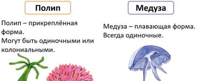
Wandering along the seashore, we often see ridges of greenish, brown or brown tangled lumps of hard threads thrown out by the waves. Very few people know that a significant part of this "sea grass" is not of vegetable, but of animal origin. Anyone who has been to the sea, of course, has seen that all the stones, piles and other underwater objects are overgrown with some kind of tender bushes wriggling in the waves. If you collect such bushes and look at them under a microscope, then along with real algae, you can see something very special. Here we have a brown jointed twig with pink lumps at the ends. At first, pink lumps are motionless, but after they stand still for a few minutes, they begin to move, stretch out in length, taking the form of a small jug with a rim of tentacles at the upper end of the body. These are hydroid polyps. eudendrium(Eudendrium), living in our northern seas, in the Black Sea and in the seas on Far East. Nearby is another, also jointed, but lighter branch. The polyps on it are also pink, but shaped like a spindle. The tentacles sit on the body of the polyp without any order, and each is equipped at the end with a small head - an accumulation of stinging cells. The movements of polyps are slow, they either bend their body, or slowly sway from side to side, but more often they sit motionless, spreading their tentacles wide - they lie in wait for prey. On some polyps, buds or young developing jellyfish can be seen. The grown-up jellyfish vigorously squeeze and unclench their umbrella, the thin thread connecting the jellyfish with the polyp breaks, and the jellyfish swims away in jerks. These are polyps Korine(Coryne) and their jellyfish. They also live in arctic and temperate seas.
And here is another bush, polyps on it sit inside transparent bells. Outwardly, they are very similar to eudendrium polyps, but behave completely differently. It is worth touching the polyp lightly with the end of the needle, as it is rapidly drawn into the depths of its protective shell, the bell. Jellyfish can also be found on the same bush: they, like polyps, are hidden inside a transparent protective shell. Jellyfish sit tightly on a thin, tentacleless polyp. It's a hydroid colony obelia(Obelia).
Now that we can distinguish hydroids from algae, we should pay attention to the feather-like colony. aglaophenia(Aglaophenia). In this species, which is very common in our Black Sea, feeding polyps sit on a branch in one row. Each is enclosed in a cup - gidroteka and surrounded by three protective polyps.
Aglaophenia does not form free-floating jellyfish, and underdeveloped individuals of the medusoid generation are hidden inside a very complex formation - a basket (a modified branch of the colony).
Colonies of hydroids settle most often at shallow depths - from the littoral to 200-250 m and prefer rocky soil or are attached to various wooden and metal objects. Often they grow very densely on the underwater parts of ships, covering them with a shaggy coat. In these cases, hydroids bring significant harm to navigation, since such a "fur coat" sharply reduces the speed of the vessel. There are many cases when hydroids, settling inside the pipes of the sea water supply, almost completely closed their gap and prevented the supply of water. It is rather difficult to fight hydroids, since these animals are unpretentious and develop quite well, it would seem, in adverse conditions. In addition, they are characterized by rapid growth - bushes 5-7 cm tall grow in a month. To clear the bottom of the ship from them, you have to put it in a dry dock. Here the ship is cleared of overgrown hydroids, polychaetes, bryozoans, sea acorns and other fouling animals.
AT recent times began to use special poisonous paints - the underwater parts of the ship covered with them are subject to fouling to a much lesser extent.
Hydroids that settle in the littoral zone are not at all afraid of the surf. In many of them, polyps are protected from blows by a skeletal cup - theca; on colonies growing in the surf zone itself, the theca is always much thicker than in the same species living deeper, where surf waves are not felt (Fig. 159).

In other hydroids from the surf zone, the colonies have a long, very flexible trunk and branches, or they are divided into segments. Such colonies meander along with the waves and therefore do not break or tear.
At great depths, special hydroids live, not similar to littoral species. Herringbone or feather-shaped colonies predominate here, many look like trees, and there are species resembling a brush. They reach a height of 15-20 cm and cover the seabed with dense forest. In the thickets of hydroids live worms, mollusks, crustaceans, echinoderms. Many of them, such as sea goat crustaceans, find refuge among hydroids, others, such as sea "spiders" (multi-legged), not only hide in their thickets, but also feed on hydropolyps.
If you move around the settlements of hydroids with a small-meshed net or, even better, use a special, so-called plankton net for this, then among the mass of small crustaceans and larvae of various other invertebrates, hydroid jellyfish will come across. Most types of hydrojellyfish are not very large animals, rarely they reach more than 10 cm in diameter of the umbrella, usually the size of hydrojellyfish is 2-3 cm, and often only 1-2 mm. Hydroid jellyfish are very transparent. You won’t even notice the jellyfish caught and placed in a glass dish right away: only whitish strings of channels and a mouth proboscis are visible. Only if you look closely, you can see the contours of the umbrella.
Looking at a hydroid colony Korine(Sogupe), we have already seen just budding small jellyfish of this species. A fully formed jellyfish has a bell-shaped umbrella 1-8 cm tall, four tentacles and a long, worm-like oral proboscis. With sharp contractions of the umbrella, the jellyfish quickly moves in a horizontal plane or rises up. Down it slowly sinks under the influence of gravity, frozen in the water with spread tentacles. Marine planktonic crustaceans, which make up the main food of the jellyfish, constantly make vertical movements: during the day they sink into the depths, and by night they rise to the surface. They descend into deeper, calmer layers of water also during waves. Jellyfish constantly move after them, two senses help them to pursue their prey - touch and sight. In calm water, the jellyfish's umbrella contracts rhythmically all the time, raising the animal to the surface. As soon as the jellyfish begins to feel the movement of water caused by the waves, its umbrella stops contracting and it slowly sinks into the depths. She distinguishes light with the help of eyes located at the base of the tentacles. Too bright light acts on it like excitement - the umbrella stops contracting and the animal plunges into a darker depth. These simple reflexes help the jellyfish to pursue prey and escape from the excitement that is fatal to it.
As mentioned above, Korine's jellyfish feeds on planktonic organisms, mainly copepods. The eyes of a jellyfish are not so perfect that she can see her prey, she catches it blindly. Its tentacles can stretch very significantly, surpassing the height of an umbrella by dozens of times. The entire surface of the tentacle is dotted with numerous stinging cells. As soon as a crustacean or some other small planktonic animal touches the tentacle, it is immediately affected by stinging cells.
At the same time, the tentacle quickly contracts and pulls the prey to the mouth. The long proboscis extends towards the prey. If a larger crustacean is caught, the jellyfish braids it not with one, but with two, three or all four tentacles.
Jellyfish with a flat umbrella and numerous tentacles catch their prey in a completely different way, for example thiaropsis(Tiaropsis) - a hydrojellyfish the size of a two-penny coin, very common in our northern seas. Along the edges of her umbrella are up to 300 thin tentacles. In a resting jellyfish, the tentacles are widely spaced and cover a significant area. When the umbrella is contracted, the jellyfish, as it were, sweeps away the crustaceans with them, fitting them to the middle of the underside of the umbrella (see Fig. 160). The mouth of the thiaropsis is wide, equipped with four large fringed lobes, with which the jellyfish captures fitted crustaceans.

Despite their small size, hydroid jellyfish are very voracious. They eat a lot of crustaceans and therefore are considered harmful animals - competitors of plankton-eating fish. Plentiful food is necessary for jellyfish for the development of reproductive products. Swimming, they scatter a huge number of eggs into the sea, which subsequently give rise to the polypoid generation of hydroids.
Above, we called the coelenterates typical inhabitants of the sea. This is true for 9000 species belonging to this type, but about one and a half to two dozen species of coelenterates live in fresh waters is no longer found in the seas. Apparently, their ancestors moved to fresh waters a very long time ago.
It is very characteristic that all these forms of both freshwater and brackish-water basins refer only to hydroid class and even only to one subclass - hydroid(Hydroidea).
Among all other coelenterates, no tendency to low salinity water is observed.
The most typical inhabitants of the fresh waters of the entire globe, often forming very dense populations, include several species hydra, constituting detachment of hydra(Hydrida).
FRESHWATER HYDRA
In each group of the animal kingdom there are representatives beloved by zoologists, whom they use as the main objects in describing the development and structure of animals and on which numerous experiments in physiology are performed. In the type of coelenterates, the hydra serves as such a classic object. This is understandable. Hydra is easy to find in nature and relatively easy to maintain in the laboratory. They multiply rapidly, and therefore in a short time it is possible to obtain mass material. Hydra is a typical representative of intestinal animals standing at the base of the evolutionary tree of multicellular organisms. Therefore, it is used in clarifying all issues related to the study of anatomy, reflexes and the behavior of lower multicellular organisms. This, in turn, helps to understand the origin of higher animals and the evolution of their physiological processes. In addition, the hydra serves as an excellent object in the development of such general biological problems as regeneration, asexual reproduction, digestion, axial physiological gradient, and much more. All this makes it an indispensable animal for both educational process- from high school to senior courses of the university, and in a scientific laboratory, where the problems of modern biology and medicine are solved in their various branches.
The first person who saw the hydra was the inventor of the microscope and the greatest naturalist of the 17th-18th centuries. Anton Levenguk.

Looking at aquatic plants, Leeuwenhoek saw among other small organisms a strange animal with numerous "horns". He also observed the growth of kidneys on his body, the formation of tentacles in them and the separation of the young animal from the mother's body. Leeuwenhoek depicted a hydra with two kidneys, and also drew the tip of its tentacle with stinging capsules, as he saw it under his microscope.
However, Levenguk's discovery almost did not attract the attention of his contemporaries. Only 40 years later, the hydra became interested in connection with the extraordinary discovery young teacher Trambley. In his spare time, studying little-known then aquatic animals, Tremblay discovered a creature that looked like both an animal and a plant. To establish its nature, Tremblay cut this creature in half. The regenerative abilities of lower animals were still almost unknown at that time, and it was believed that only plants could restore lost parts. To Tremblay's surprise, a whole hydra grew from each half, both of them moving, grabbing prey, which means it was not a plant. The possibility of turning a piece of the body of a hydra into a whole animal was perceived as a significant discovery in the science of life, and Tremblay engaged in a deep and serious study of the hydra. In 1744, he published the book "Memoirs on the History of a Genus of Freshwater Polyps with Horned Arms." The book described in great detail the structure of the hydra, its behavior (movements, catching prey), reproduction by budding, and some aspects of physiology. To test his assumptions, Tremblay did a series of experiments with the hydra, laying the foundation for a new science, experimental zoology.
Despite the imperfection of the then optics and the poor development of zoology, Tremblay's book was written in such a high scientific level, which has not lost its significance to this day, and the drawings from this book can be found in many textbooks on zoology.
Now the scientific literature on the hydra amounts to many hundreds of articles and books, but nevertheless, the hydra still occupies the minds of researchers. A small primitive animal serves as a touchstone for them, on which many issues are resolved. modern science about life.
If you collect aquatic plants in the coastal part of a lake or river and place them in an aquarium with clean water, then soon you can see hydras on them. At first they are almost invisible. Animals that are disturbed shrink strongly, their tentacles contract. But after some time, the body of the hydra begins to stretch, its tentacles lengthen. Now the hydra can be properly seen. The shape of her body is tubular, at the front end there is a mouth opening, surrounded by a corolla of 5-12 tentacles. Immediately below the tentacles, the hydras of most species have a slight narrowing - a neck that separates the "head" from the body. The posterior end of the hydra is narrowed into a more or less long stalk, or stalk, with a sole at the end (in some species, the stalk is not expressed). In the middle of the sole there is a hole, the so-called aboral pore. The gastric cavity of the hydra is solid, there are no partitions in it, the tentacles are hollow, finger-like gloves.
The wall of the body of the hydra, like that of all coelenterates, consists of two layers of cells, their fine structure has already been described above, and therefore here we will focus on only one feature of the cells of the hydra body, which has been fully studied so far only on this object and has not been found in others. coelenterates.
The structure of the ectoderm (and endoderm) in different parts of the body of the hydra is not equivalent. So, at the head end of the ectoderm cells are smaller than on the body, there are fewer stinging and intermediate cells, but a sharp boundary between the integuments of the “head” and the body cannot be drawn, since the change in the ectoderm from the body to the “head” occurs very gradually. The ectoderm of the sole of the hydra consists of large glandular cells; at the point of transition of the sole to the stalk, the glandular character of the integumentary cells is gradually lost. The same can be said about the cells of the endoderm. Digestive processes occur in the middle part of the body of the hydra, here its endoderm has a large number of digestive glandular cells, and the epithelial-muscular cells of the endoderm of the middle part of the body form numerous pseudopodia. Digestion of food does not occur in the head section of the gastric cavity, in the stalk and in the tentacles. In these parts of the body, the ectoderm has the appearance of a lining epithelium, almost devoid of digestive glandular cells. Again, a sharp boundary between the cells of the digestive section of the gastric cavity, on the one hand, and such cells of the "head", stalk and tentacles, on the other hand, cannot be drawn.
Despite the difference in the structure of the cell layers in different parts of the body of the hydra, all of its cells are not in strictly defined permanent places, but are constantly moving, and their movement is strictly regular.
Using the high ability of hydra to heal wounds, you can do this interesting experience. They take two hydras of the same size and one of them is stained with some kind of vital paint, that is, with such a dye that penetrates into the tissues of the hydra without killing it. Usually, a weak aqueous solution of nilblausulfate is used for this, which stains the tissues of the hydra blue. After that, the hydras undergo an operation: each of them is cut into three parts in the transverse direction. Then, the head and lower ends of the unpainted specimen are added to the middle part of the "blue" hydra. The sections quickly fuse with each other, and we get an experimental hydra with a blue belt in the middle of the body. Shortly after the operation, one can observe how the blue belt spreads in two directions - towards the head end and the stalk. At the same time, it is not the paint that moves along the body of the hydra, but the cells themselves. The layers of ectoderm and endoderm seem to “flow” from the middle of the body to its ends, while the nature of their constituent cells gradually changes (see Fig. 162).

In the middle part of the body of the hydra, the cells multiply most intensively, and from here they move in two opposite directions. Thus, the composition of the cells is constantly updated, although outwardly the animal remains almost unchanged. This feature of the hydra is of great importance in solving questions about its regenerative abilities and for assessing data on lifespan.
Hydra is a typical freshwater animal, only in very rare cases, hydras were found in slightly saline water bodies, for example, in the Gulf of Finland Baltic Sea, and in some brackish lakes, if the salt content in them did not exceed 0.5%. Hydras live in lakes, rivers, streams, ponds, and even ditches, as long as the water is clear enough and contains high amounts of dissolved oxygen. Hydras usually keep near the coast, in shallow places, as they are photophilous. When keeping hydras in an aquarium, they always move to its illuminated side.
Hydras are sedentary animals, most of the time they sit in one place, attaching their soles to a branch of an aquatic plant, a stone, etc. Hydra's favorite posture in a calm state is to hang upside down with its tentacles lowered somewhat apart.
The hydra is attached to the substrate due to the sticky secretions of the glandular cells of the ectoderm of the sole, and also using the sole as a sucker. The hydra is held very firmly, it is often easier to break it than to separate it from the substrate. If you watch a sitting hydra for a long time, you can see that its body is slowly swinging all the time, describing a circle with its front end. Hydra can arbitrarily leave the place it sits very quickly. At the same time, apparently, it opens the aboral pore located in the middle of the sole, and the suction action stops. Sometimes you can watch the hydra "walk". First, she bends the body to the substrate and strengthens on it with the help of tentacles, then pulls the posterior end and strengthens it in a new place. After the first “step”, he takes the second, and so on, until he stops at a new place.

Thus, the hydra moves relatively quickly, but there is another, much slower, way of moving - sliding on the sole. By the force of the muscles of the sole, the hydra barely noticeably moves from place to place. It takes a very long time to notice the movement of the animal. Hydras can swim in the water column for some time. Having detached from the substrate and spreading its tentacles widely, the hydra falls to the bottom very slowly, it is able to form a small gas bubble on the sole, which drags the animal upward. However, hydras rarely use these modes of locomotion.
Hydra is a voracious predator, it feeds on ciliates, planktonic crustaceans, low-bristle worms, and also attacks fish fry. Hydras lie in wait for their prey, hanging on some twig or stem of an aquatic plant, and, spreading their tentacles widely, constantly make circular search movements. As soon as one of the tentacles of the hydra touches the victim, the rest of the tentacles rush to it and paralyze the animal with stinging cells. Now there is no trace of the slowness of the hydra, it acts quickly and "decisively". The prey is pulled by tentacles to the mouth and quickly swallowed. Hydra swallows small animals whole. If the victim is somewhat larger than the hydra itself, it can also swallow it. At the same time, the mouth of the predator opens wide, and the walls of the body are strongly stretched. If the prey does not fit into the gastric cavity as a whole, the hydra swallows only one end of it, pushing the victim deeper and deeper as it digests. A well-fed hydra shrinks somewhat, and its tentacles contract.
In the gastric cavity, where digestive processes are just beginning, the reaction of the medium is slightly alkaline, and in the digestive vacuoles of the endoderm, where digestion ends, it is slightly acidic. Hydra can absorb fats, proteins and animal carbohydrates (glycogen). Starch and cellulose, which are of plant origin, are not absorbed by hydra. Undigested food remains are expelled through the mouth.
Hydras reproduce in two ways: vegetatively and sexually. Vegetative reproduction in hydras is budding. The buds arise in the lower part of the body of the hydra above the stem, the subsequent buds are slightly higher than the previous ones, sometimes they sit on opposite sides of the hydra body, sometimes they are arranged in a spiral (the order of appearance and location of the buds depends on the type of hydra). At the same time, 1 - 3, rarely more buds develop on the body of the hydra, however, hydras with 8 or more buds were observed.

At the first stages, the kidney appears as a barely noticeable conical tubercle, then it is pulled out, taking on a more or less cylindrical shape. At the outer end of the kidney, rudiments of tentacles appear, at first they look like short blunt outgrowths, but gradually stretch out, and stinging cells develop on them. Finally, the lower part of the body of the kidney thins into a stalk, and a mouth opening breaks through between the tentacles. The young hydra still remains connected to the mother's organism for some time, sometimes the next generation of buds are even laid on it. The separation of budding hydras occurs in the same sequence in which buds arise. The young hydra is somewhat smaller than the parent hydra and has an incomplete number of tentacles. Missing tentacles appear later.
After abundant budding, the mother hydra is depleted and no buds appear on it for some time.
Some researchers also observed the division of hydras, but this method of reproduction, apparently, should be classified as abnormal (pathological) processes. Hydra division occurs after damage to its body and can be explained by the high regenerative capacity of this animal.
With abundant nutrition, the entire warm period of the year, hydras reproduce by budding, they begin sexual reproduction with the onset of autumn. Most types of hydra are dioecious, but there are also hermaphrodites, that is, those in which both male and female germ cells develop on the same individual.

Gonads are formed in the ectoderm and look like small tubercles, cones or rounded bodies. The order of appearance and the nature of the location of the gonads are the same as those of the kidneys. Each female gonad produces one egg.
In the developing gonads, a large number of intermediate, undifferentiated cells accumulate, from which both future germ cells and "nourishing" cells are formed, due to which the future egg grows. At the first stages of egg development, intermediate cells acquire the character of mobile amoeboids. Soon one of them begins to absorb the others and increases significantly in size, reaching 1.5 mm in diameter. After that, a large amoeboid picks up its pseudopodia and its outlines are rounded. Following this, two divisions of maturation occur, in which the cell is divided into two unequal parts, and on the outer side of the egg there remain two small so-called reduction bodies - cells separated from the egg as a result of division. At the first division of maturation, the number of egg chromosomes is halved. The mature egg emerges from the gonad through a gap in its wall, but remains connected to the body of the hydra with the help of a thin protoplasmic stalk.
By this time, spermatozoa develop in the testes of other hydras, which leave the gonad and swim in the water, one of them penetrates the egg, after which cleavage immediately begins.
At the time when the cells of the developing embryo are dividing, on the outside it is dressed in two shells, the outer of which has rather thick chitinoid walls and is often covered with spines. In this state, the embryo, under the protection of the double shell-embryotheca, overwinters. (Adult hydras die with the onset of cold weather.) By spring, there is already an almost formed small hydra inside the embryotheca, which leaves its winter shell through a break in its wall.
Currently, about a dozen species of hydras are known that inhabit the fresh waters of the continents and many islands. Different types of hydras differ from each other very slightly. One of the species is characterized by a bright green color, which is due to the presence in the body of these animals of symbiotic algae - zoochlorella. Among our hydras, the most famous stalked, or brown, hydra(Hydra oligactis) and stemless, or - ordinary, hydra(Hydra vulgaris).
How does the hydra behave in its environment, how does it perceive irritations and respond to them?
Like most other intestinal cavities, hydra responds to any adverse irritation with a contraction of the body. If the vessel in which the hydras are sitting is slightly shaken, then some of the animals will shrink immediately, such a push will not affect others at all, some of the hydras will only slightly tighten their tentacles. This means that the degree of reaction to irritation in hydras is very individual. Hydra is completely devoid of the ability to "remember": you can prick it for hours with a thin pin, but after each contraction it is again pulled in the same direction. If the injections are very frequent, then the hydra stops responding to them.
Although hydras do not have special organs for the perception of light, they definitely react to light. The anterior end of the hydra is most sensitive to light rays, while its stalk almost does not perceive light rays. If you shade the whole green hydra, then it will shrink in 15-30 seconds, but if you shade the headless hydra or shade only the stalk of the whole hydra, then it will shrink only after 6-12 minutes. Hydras are able to distinguish the direction of the flow of light and move towards its source. The speed of movement of the hydra towards the light source is very low. In one of the experiments, 50 green and the same number of brown hydras were placed in a vessel at a distance of 20 cm from the glass wall through which the light fell. The first to move towards the light were the green hydras; after 4 hours, 8 of them reached the light wall of the aquarium, after 5 hours there were already 21 of them, and after 6 hours - 44. By this time, the first 7 brown hydras also came there. In general, it turned out that brown hydras went into the light worse, only after 10 hours 39 brown hydras gathered at the light wall. The rest of the experimental animals were still on the way by this time.
The ability of hydras to move towards a light source or simply move to lighter areas of the pool is very important for these animals. Hydras feed mainly on planktonic crustaceans - cyclops and daphnia, and these crustaceans always stay in bright and well-warmed places by the sun. Thus, going towards the light, hydras approach their prey.
For a researcher studying the reactions of lower organisms to light, hydras open up the widest field of activity. You can set up experiments to identify how sensitive animals are to weak or, on the contrary, very strong light sources. It turned out that hydras do not react at all to too weak light. Very strong light makes the hydra go to shaded places and can even kill the animal. Experiments were carried out to determine how sensitive the hydra is to changes in light intensity, how it behaves between two light sources, and whether it distinguishes between individual parts of the spectrum. In one of the experiments, the wall of the aquarium was painted in all colors of the spectrum, while green hydras gathered in the blue-violet region, and brown ones in the blue-green region. This means that hydras distinguish color, and their different species have different “tastes” for it.
Hydras (except green) do not need light for normal life. If they are well fed, they live well in the dark. The green hydra, in whose body symbiotic zoochlorella algae live, even with an abundance of food in the dark, feels bad and is greatly reduced.
On hydras, experiments can be carried out on the effects on the body of various kinds of harmful radiation. So, it turned out that brown hydras die after a minute of exposure to ultraviolet rays. The green hydra turned out to be more resistant to these rays - it dies only at the 5-6th minute of exposure.
Experiments on the effect of X-rays on hydras are very interesting. Small doses of X-rays cause increased budding in hydras. Irradiated hydras, compared with non-irradiated ones, give about 2.5 times more offspring in the same period. Increasing the dose of radiation causes suppression of reproduction; if the hydras receive too much X-rays, they die soon after. It is important to note that low doses of radiation increase the regenerative abilities of hydro.
When exposed to radioactive radiation, a completely unusual result was obtained. It is well known that animals do not feel radioactive rays in any way and therefore, once in their zone, they can receive a lethal dose and die. The green hydra, reacting to the radiation of radium, seeks to get away from its source.
From the above examples, it can be seen that such experiments with hydras, such as studying the influence of various environmental factors on them, are not empty fun, not science for the sake of science, but a serious and very important matter, the results of which can give very significant practical conclusions.
Of course, the effect on the hydra of temperature, the concentration of carbon dioxide, oxygen, as well as a number of poisons, drugs, etc., was studied.
Hydra proved to be a very convenient object for carrying out a number of experimental studies on the study of the phenomenon of regeneration in animals.
As has been repeatedly mentioned, hydra easily restores lost body parts. The animal, cut in half, soon restores the missing parts. But it becomes unclear: why does a “head” with tentacles always grow at the front end of the segment, and a stalk at the back? What laws govern recovery processes? It is likely that some of these laws may be common to both the hydra and the more highly organized animals. Once you know them, you can important findings applicable even to medicine.
It is very easy to perform operations on hydras, it does not require any anesthetics or complex surgical instruments. All the equipment of the “operating room” consists of a needle inserted into a wooden handle, a sharp eye scalpel, small scissors and thin glass tubes. The first experiments to determine the regenerative abilities of the hydra were carried out more than 200 years ago by Tremblay. This painstaking researcher observed how whole animals arise from the longitudinal and transverse halves of hydras. Then he began to make longitudinal incisions and saw that stems were formed from the shreds in the lower part of the polyp, and “heads” were formed from the shreds in its upper part. Repeatedly operating on one of the experimental polyps, Tremblay received a seven-headed polyp. Having cut off all seven “heads” for him, Tremblay began to wait for the results and soon saw that a new one appeared in place of each cut off “head”. The seven-headed polyp, in which severed “heads” grow again, was like two drops of water similar to a mythical creature - the Lernean hydra, slain by the great hero ancient greece Hercules. Since then, the name hydra has been preserved behind the freshwater polyp.
Along the way, Tremblay found that the hydra is restored not only from halves, but also from very small pieces of the body. Now it has been established that even from 1/200 of the body part of the hydra, a whole polyp can develop. However, later it turned out that the regenerative ability of such small pieces from different parts of the hydra's body is not the same. A section of the sole or stalk is restored into a whole hydra much more slowly than a section from the middle part of the body. However, this fact remained unexplained for a long time.
The internal forces regulating and directing the processes of normal regeneration were discovered much later by the famous American physiologist Child (CM. Child). Child found that in a number of lower animals there is a pronounced physiological polarity in the body. So, under the influence of toxic substances, cells on the body of an animal die and are destroyed in a very definite sequence, namely from the front end to the back (in hydra from the “head” to the “sole”). Therefore, the cells located in different parts of the body are physiologically unequal. The difference between them lies in many other manifestations of their physiology, including the effect on developing young cells at the site of injury.
The gradual change in the physiological activity of cells from one pole to another (along the axis of the body) is called the axial physiological gradient.
Now it becomes clear why the pieces cut from the sole of the hydra are very slowly restored by the hypostome and tentacles - the cells that form them are physiologically very far from the cells that form the "head". The axial gradient plays a very important role in regeneration, but other factors also have a noticeable influence on this process. During regeneration, the presence on the regenerating part of a developing kidney or an artificially transplanted area of tissue from another part of the animal's body, especially from its front part, is of great importance. Possessing high physiological activity, the developing kidney or cells of the "head" in a certain way affect the growth of regenerating cells and subordinate their development to their influence. Such groups of cells or organs that make their own adjustments to the action of the axial gradient are called organizers. The elucidation of these features of regeneration helped to understand many obscure questions in the development of the animal organism.
AT largest center physiology - in the institute created by Academician Pavlov in Koltushi there is a monument to a dog. Most of laws set forth in the teachings of Pavlov, was discovered when setting up experiments on dogs. Perhaps a small freshwater polyp deserves the same monument.
FRESHWATER MEDUSA
In 1880, jellyfish suddenly appeared in the pool with tropical plants of the London Botanical Society. Immediately two zoologists Lankester (Lankester) and a major connoisseur of coelenterates Olmen (A1man) reported this find in the pages of the journal "Nechur" ("Nature"). The jellyfish were very small, the largest of them barely reaching 2 cm in diameter of an umbrella, but their appearance excited the then zoologists: before that, they had not imagined that freshwater jellyfish could exist. Jellyfish were considered typical inhabitants of the sea. Shortly before this, the magnificent South American aquatic plant Victoria Regia had been planted in the pool, so it has been suggested that the jellyfish were brought to London along with planting material from the Amazon. After a while, the jellyfish disappeared from the pool as mysteriously as they appeared. They were discovered again only five years later, also in London, but in a different pool with the same tropical plant. In 1901, these jellyfish appeared in Lyon (France), also in the greenhouse pool with Victoria Regia. Then they began to be found in Munich, Washington, St. Petersburg, Moscow. Jellyfish were found either in the pools of botanical gardens or in aquariums with tropical fish. To the surprise of amateur aquarists, they suddenly have new pets. Tiny jellyfish (often only 1 - 2 mm in diameter of an umbrella) suddenly appeared in large numbers in an aquarium in which there had not been a single one the day before. For several days it was possible to observe how the jellyfish move in jerks in the water and willingly eat small crustaceans. But one fine day, looking into his aquarium, the owner found only fish in it, there were no jellyfish there.
By this time, the freshwater jellyfish had been described in detail in specialized zoological literature. It turned out that she belonged to hydroid class. They called her kraspedakustoy(Craspedacusta). The smallest jellyfish have a hemispherical umbrella, 4 radial canals and 8 tentacles. As the jellyfish grows, the shape of its umbrella becomes more and more flat, and the number of tentacles increases.

Sexually mature jellyfish reach 2 cm in diameter and carry a wide sail along the edge of the umbrella and about 400 thin tentacles seated with stinging cells. The oral proboscis is tetrahedral, with a cruciform mouth opening, the edges of the mouth are slightly folded. At the point of origin of the radial canals from the oral proboscis, 4 gonads develop. Jellyfish are very transparent, their mesoglea is colorless, and their tentacles, radial canals, oral proboscis and gonads are whitish or cream in color.
This jellyfish made a wish to zoologists difficult riddle. If we agree with the opinion that it gets into greenhouses along with plants from the tropics, then how does it survive during transportation? Victoria-regia was transported from the shores of the Amazon in the form of seeds or rhizomes. Delicate jellyfish, accidentally captured along with rhizomes, must surely die during the long journey across the ocean. But even if we assume that the jellyfish, despite drying out, can survive, then how does it get into small aquariums of exotic fish lovers?
Soon, jellyfish began to be found in natural reservoirs. The first time she was caught in the Yangtze River in China, then in Germany, then in the USA. However, both in natural and artificial reservoirs, the finds were very rare and always unexpected: for example, once a jellyfish was found in the vaults of the Washington water supply.

Observations on the jellyfish made it possible to establish that it buds FROM tiny tentacleless polyps, called microhydras(Microhydra). These polyps were found back in 1884 in the same pools in London where jellyfish were also caught, but then no one suspected a connection between these two such dissimilar creatures. Microhydra polyps are visible to the naked eye as white dots against the background of green leaves of aquatic plants on which they usually settle. Their height usually does not exceed 0.5-1 mm, the shape of the body resembles a skittle: the body is in the form of a bottle, and a spherical “head” with a mouth in the middle sits on a short neck. The head is densely seated with stinging cells, there are no tentacles. Polyps sometimes form primitive colonies of 2-7 individuals. Microhydra reproduces by budding and forms tentacleless polyps similar to itself. From time to time, a group of cells shaped like a small worm separates from one side of the polyp's body. Such groups of cells are called frustules. Frustula is able, wriggling, to crawl along the bottom and climb onto aquatic plants, here it turns into a young microhydra.
Once it was possible to observe how a jellyfish began to develop on the body of a microhydra from a kidney; when she separated from the polyp and began to swim, it was easy to recognize a young craspedacusta in her. It was also possible to follow the development of craspedacusta eggs. Initially, a worm-like larva is formed from the egg, devoid of cilia and very similar to the microhydra frustula. After some period of crawling along the substrate, the larva attaches to it and turns into a tentacleless polyp. Thus, it was established that the jellyfish kraspedakusta and the polyp microhydra belong to the same species of coelenterates, but to its different generations.
The experiments carried out showed that at the change of generations this type of hydroid is extremely big influence provided by environmental conditions. Budding of jellyfish on polyps occurs only at a water temperature not lower than 26-33°C, and budding of polyps and separation of frustules occurs at a temperature of 12-20°C. After that, it became clear that the existence of a species can be maintained for a long time due to the reproduction of polyps. Neither aquarists nor botanists in greenhouses pay attention to small motionless microhydras, since they are almost invisible to the naked eye, it is very difficult to find them in nature. Polyps can live for a long time in an aquarium, and when the temperature rises, all polyps develop jellyfish buds and they separate jellyfish. Kraspedakusta jellyfish are mobile and can be seen in the water with the naked eye. Now it becomes clear why they were almost always found in pools with tropical plants and fish: these pools were artificially heated. Only one thing is unclear: have jellyfish always lived in Europe or were they brought there? (Polyps may be able to tolerate some desiccation and a long journey in adverse conditions.) And where is the homeland of microhydra-craspedacusta?
It is rather difficult to answer this question. Since the first discovery of jellyfish in London, over 100 cases of their presence in various parts of the world have been described. Here short description distribution of the species. In the USSR, their habitat is the Lyubov reservoir near Tula, the Don River, Lake Karayazi near Tbilisi (at an altitude of almost 2000 m above sea level), the Kura River, and artificial reservoirs in Old Bukhara. In addition, jellyfish and polyps have repeatedly appeared in aquariums of amateur fish farmers and at the universities of Moscow and Leningrad. Outside of our country, this species was found in almost all European countries, in India, China and Japan, in Australia, North and South America. It is impossible to indicate now where his homeland is and where he was brought.
More recently, this type of coelenterates again made zoologists think. Now that the distribution, lifestyle, structure of polyps and jellyfish seemed to be well studied, it suddenly became clear that polyps of two genera can develop from the eggs of Kraspedakusta - the tentacleless and tentacled polyps described above. Both genera of polyps form frustules. Tentacle-bearing polyps with the help of budding form similar to themselves and tentacleless polyps, they cannot bud jellyfish. Tentacleless polyps form polyps and jellyfish similar to themselves, but are not able to bud polyps equipped with tentacles. Both forms of polyps are formed from frustules. Tentacle-bearing polyps have only been discovered twice so far: in 1960 in Hungary and in 1964 in an aquarium Leningrad University. The conditions that cause their appearance are not yet clear. In the rivers of India and the great lakes of Africa, there are two more species of freshwater jellyfish, close relatives of kraspedakusta. A well-known jellyfish from the African Lake Tanganyika, called limnocnida(Limnocnida tanganjice).
ORIGIN OF FRESHWATER CELESTONES
Of these hydroids, first of all it is necessary to say about Cordylophora.

Cordylophora forms small, tender colonies in the form of bushes up to 10 cm high. Polyps sit at the ends of branches and have a spindle shape. Each polyp has 12-15 tentacles, sitting without a strict order in the middle part of the body. There are no free-swimming jellyfish in Cordylophora; individuals of the jellyfish generation are attached to the colony.
This species was first discovered by Academician of the Russian Academy P.S. Pallas in 1771 in the northern part of the Caspian Sea, because cordylophora and is called the Caspian (Cordylophora caspia). However, its distribution is by no means limited to this basin, it lives in the Baltic, Black and Azov seas, and is also found along the entire Atlantic coast of Europe and in the mouths of all major rivers Asia, America and Australia. This species settles only in heavily desalinated areas of the sea and lives at shallow depths, usually no deeper than 20 m.
The name given by Pallas to Cordylophore - Caspian - has its own meaning. The fact is that the birthplace of Cordylophora is the Caspian Sea. Only in the middle of the last century, Cordylophora penetrated the Baltic Sea along the Volga and the Mariinsky system, where, due to its low salinity (0.8%), it found its second home. Cordylophora is an organism that grows; it settles on all solid underwater objects, both fixed and moving. Further assistance in resettlement was provided by countless ships flowing from all sides into the Baltic Sea. Returning home, they took away from the Baltic Sea on their bottom an intruder, a “violator of borders”.
But how did free-living coelenterates get into fresh water bodies? Can they not use the mouths of the rivers flowing into the sea for this? Of course they can, but in doing so they have to overcome two hurdles. One of them is a decrease in salinity. Only species that can withstand very significant desalination can enter rivers.
Among typical marine life, there are those on which even the slightest decrease in the percentage of salt in sea water has a detrimental effect. Almost all of them are coral polyps, scyphoid jellyfish and most hydroids. But some hydroids can still exist even with some desalination. Of the coelenterates mentioned in this book, Corine belongs to the euryhaline. This species can live both in water with normal oceanic salinity and in desalinated seas, for example, in the White and Black.
From among the euryhaline species, those whose descendants actively made their way into freshwater reservoirs came out. The process of conquering rivers and lakes proceeded gradually. At first, a group of brackish-water hydroids stood out, which could no longer return to the ocean, since they could not stand the high salinity of its waters. Then already brackish-water came close to the mouths of the rivers. Not all of them were able to overcome this "barrier", most of them remained in the river mouth. Cordylophora is currently following this path.
Once in the river, marine animals met on their way another "barrier" - the current. With the active penetration of marine or brackish coelenterates into fresh waters, they inevitably had to overcome the oncoming flow of water, which carried planktonic jellyfish and attached polyps or their colonies incapable of independent movement back into the sea. Promotion of such attachment polyps towards the current was difficult.
In remote geological epochs, the map of the Earth was different from what we see it now. In many places, the modern land was covered by the sea. When the sea left, closed salt pools remained, and marine animals were preserved in them. Some of these pools gradually desalinated, and the animals either died or adapted to new conditions. The now closed Caspian Sea, which is essentially a huge brackish lake, was formerly connected to the ocean, and many animals of marine origin have been preserved in it. Among them is an interesting coelenterate - Pallas Merizia(Moerisia pallasi). This type of hydroid has two forms of polyps: some live in a colony at the bottom, others lead a planktonic lifestyle. Floating polyps form colonies of two individuals connected to each other by their legs. From time to time, the colony is torn in half, and at the site of the rupture, each polyp forms a new corolla, tentacle and mouth. In addition, polyps also reproduce by budding, separating small free-swimming jellyfish from themselves. One close species of merizia lives in the Black and Azov Seas, the other in the salt lakes of Northeast Africa.

It is quite clear that all three types of merisia originated from one common ancestor that once lived in the ancient Sarmatian Sea. When the Sarmatian Sea left, a number of reservoirs remained in its place, including the closed Caspian Sea and the lakes of Egypt. They developed independent types of merizia.
If you imagine that the desalination of the reservoir goes even further, then you can understand how freshwater jellyfish can arise. Their way of conquering freshwater basins is a long-term adaptation to increasing desalination. At the same time, they do not need to move anywhere, they make their way from the sea to fresh water not in space, but in time.
In 1910 on the Atlantic coast North America several small hydrojellyfish were caught. It turned out that they belong to a previously unknown species. By itself, this fact is of little importance. And now several new species of coelenterates are described annually - there is still a lot of unexplored in the sea. Another thing is interesting. This jellyfish is named blackfordia(Blackfordia) - after 15 years was caught in the Black Sea. Neither in the Mediterranean Sea, whose fauna is very well known, nor on the European coast Atlantic Ocean this species does not live. How did the American Blackfordia end up in the Black Sea? The second incident happened very recently. One of the types of hydroids that live in the Kiel Canal is bougainvillea- was unexpectedly discovered again in the Black Sea. And blackfordia and the mentioned Baltic hydroid(Bougainvillia megas) - brackish water species; in order to get from one basin with low salinity to another, they must, like Cordylophora, overcome an obstacle - the sea with its high salinity.
Before the canal was built between the Volga and the Don, there were only two types of coelenterates in the Caspian Sea - the Caspian Merizia and the Cordylophora. When the canal was ready and navigation began, three more species moved from the Azov-Black Sea basin to the Caspian Sea. A year after the canal was put into operation, blackfordia moved to the Caspian Sea, a year later the Black Sea merizia, and after it the Baltic hydroid (Bougainvillia megas), which had recently entered the Black Sea from the Kiel Bay. Of course, not only coelenterates travel this way, but also molluscs, and crustaceans, and worms, and other brackish-water organisms.
“SAILING FLEET”
Hydroid class is divided into two subclasses - hydroids and siphonophore. We now turn to the description of these amazing pelagic colonial coelenterates.
A whole world of living beings lives on the verge of two elements - water and air. On floating algae, fragments of wood, pieces of pumice and other objects, you can find a variety of adherent or firmly clinging animals. You should not think that they got here by accident - "in distress." On the contrary, many of them are closely connected with both the water and the air environment, and in other conditions they cannot exist. In addition to such "passive passengers", here you can also see animals actively swimming near the surface itself, equipped with variously arranged organs - floats, or animals that are held using a film of surface tension of water. This whole complex of organisms (pleuston) is especially rich in the subtropics and tropics, where the destructive effect of low temperatures is not felt.
Above, when it came to the action of stinging cells, the “Portuguese warship” was already mentioned - a large siphonophore physalia(Physalia, see color table 8).

Like all siphonophores, physalia is a colony, which includes both polypoid and medusoid individuals. An air bubble rises above the surface of the water, OR a pneumatophore, a modified medusoid individual of the colony. In large specimens, the pneumatophore reaches 30 cm. It usually has a bright blue or reddish color. An air bubble floats on the surface of the sea like a tightly inflated rubber balloon. The gas filling it is close in composition to air, but differs in an increased content of nitrogen and carbon dioxide and a reduced amount of oxygen. This gas is produced by special gas glands located inside the bladder. The walls of the pneumatophore can withstand rather strong gas pressure, as they are formed by two layers of ectoderm, two layers of endoderm and two layers of mesoglea. In addition, the ectoderm secretes a thin chitinoid membrane, due to which the strength of the pneumatophore also increases significantly, although its walls remain very thin. Top part The pneumatophore has an outgrowth in the form of a comb. The crest is located on the pneumatophore somewhat diagonally and has a slightly curved S-shape. All other individuals of the colony are located on the underside of the pneumatophore and are submerged in water.
Feeding polyps, or gastrozoids, sit in one row. They are more or less bottle-shaped and face down with their mouths. Each feeding polyp is equipped with one long tentacle - a noose. Throughout its length, the noose is densely covered with stinging cells. Next to each feeding polyp on the underside of the bubble is attached the base of the gonodendra - an individual of the polypoid generation. On the gonodendra and its lateral outgrowths there are clusters of reduced medusoid individuals - gonophores, in which reproductive products develop. Protective tentacleless polyps - palpons - also sit here. Each gonodendra has one medusoid, called a nektophore or swimming bell. Sex cells in the nektophore are not formed, and its umbrella reaches a significant size and is able to contract, like in free-swimming jellyfish. Before the onset of sexual maturity of the gonophores, the gonodendra break away from the colony and float near the sea surface, with the nectophore performing locomotor functions.

Due to the oblique arrangement of the ridge on the swim bladder, the physalia is asymmetric, and two forms of physalia are known - “right” and “left”, which are, as it were, a mirror image of each other. It was noticed that all physalians living in one part of the sea have the same structure, that is, they are all either “right” or “left”. In this regard, it has been suggested that there are two species or two geographical races of Physalia.
However, when they began to study the development of these siphonophores, it was found that among the offspring of one physalia there is always an equal number of both “right” and “left”. This means that there are no special races for physalia. But how do clusters of "left" and "right" siphonophores arise, and why do these two forms not occur together?
The answer to this question was obtained after a detailed study of the structure of the air bladder of physalia. It turned out that the shape and location of the crest at its top are very important for physalia. As mentioned above, the crest of the physalia is slightly curved in the shape of the letter S. The physalia moves along the surface of the sea due to the fact that the wind hits its air bubble. If there were no ridge, the siphonophore would constantly move in a straight line and would eventually be washed ashore. But the presence of a crest makes significant changes to the sailing equipment of the "Portuguese boat". An obliquely set and curved crest makes the animal swim at an acute angle to the wind and from time to time make a turn around its axis against the wind.
If you observe a physalia floating near the shore, towards which the wind is blowing, you can see how it either approaches the shore, then, unexpectedly turning to the observer on the other side, slowly sails away from him. Entire armadas of “Portuguese ships” maneuver in this way, reminiscent of the actions of the sailing fleet of the period of medieval wars. When moving, the "right" and "left" "Portuguese boats" behave differently. Under the influence of the wind blowing in one direction, they diverge in different directions - "right" to the left, and "left" to the right. That's why there are clusters of identical forms of physalia.
Pleistonic organisms also include very peculiar coelenterates - porpita(Porpita) and velella(Velella), which is also called a sailboat.
For a long time, these animals were classified as siphonophores, and their individual appendages were considered specialized individuals of the colony. Now more and more zoologists are inclined to believe that the porpita and the sailboat are not a colony, but a large single floating polyp, and classifies them as order chondrophore(Chondrophora) of hydroid class. Their body is flattened; in porpita it has the shape of a circle, in a sailboat it is oval. The upper side of the disc is covered with a chitinoid membrane, under which is placed a complex air bell - pneumatophore. It consists of a central chamber, a large number of annular chambers surrounding it and thin tubes extending from them to all parts of the body - tracheas, which are used for breathing. On the underside of the disk are the organs of the polyp. There is a mouth cone in the center, and numerous tentacles are located along the periphery. Between the mouth cone and the tentacles there are special outgrowths of the body - gonodendria, on which individuals of the medusoid generation bud. The upper side of the coastal porpita disc is smooth; a velella living in the open ocean has a high triangular outgrowth on it - a sail. The sail of the velella has the same meaning as the crest on the air bladder of the physalia. It is located asymmetrically and slightly S-shaped on the oval body of the sailboat. The sail allows the animal to move not in a straight line, but to maneuver, although, of course, not arbitrarily, but more or less randomly.
In the subtropical parts of the ocean, where the temperature does not fall below 15 ° C, sailboats are found in very large numbers. In some places, these large coelenterates (they reach 12 cm along the long axis of the disk) gather in huge flocks several tens of miles long, and for each square meter The surface of the ocean is accounted for by the sailboat. Together with large sailboats, young ones also swim, the size of which is measured in millimeters.
The wind, striking the sail, drives a flock of velles across the sea, and they can travel many hundreds of miles.
Living in the open ocean, sailboats are not afraid of water: they cannot drown, as they have a very perfect pneumatophore, consisting of a large number of independent chambers. If the wave nevertheless overturns the velella, then with the help of movements of the edges of the disk, it assumes a normal position and again exposes the sail to the wind. In addition to sailboats, you can also meet many other animals here, which, however, are almost invisible at first.
It is well known that the high seas of the tropics have an intense blue color. In this regard, sailboats and most of the animals that live with them are also painted in blue or blue tones - this serves as a good protection for them.
Sailboats and other animals living among them create a special, closely connected little world in the open sea - a pleistonic biocenosis, which, by the will of the current and wind, floats all the time on the surface of the ocean.
Velella, like all coelenterates, is a predator; it feeds on plankton, its food includes crustaceans, larvae of various invertebrates, and fish fry. All other animals that are part of the floating biocenosis either feed on sailboats or use them as a permanent or temporary substrate for attachment. Thus, the entire biocenosis exists due to plankton, but only sailboats directly use plankton.
On the upper side of the velella disk, as on the deck of a ship, small blue crabs travel planes(Planes). Here they find protection from enemies, and also get food. A hungry crab quickly moves to the underside of the sailboat disk and takes away the captured planktonic crustaceans from it. Having eaten, the crab again climbs onto the upper side of the disk and settles under the sail, closely clinging to it. Crabs never devour their ship, which is not the case with many other pleustian animals.
On the underside of the sailboat, you can often find the predatory gastropod mollusc Janthina. Yantins eat soft tissues until only a chitinoid skeleton remains from the sailboat. Having lost its support, the yantina does not sink, as it is well adapted to life near the surface of the water. As soon as the eaten sailboat begins to sink, the yantin releases abundant mucus, which forms air-filled bubbles. This mucus hardens very quickly, and a good float is obtained, on which the mollusk can swim independently, moving from one sailboat to another. Swim to new victim, the yantina leaves the now unnecessary float and quickly crawls onto the velella. An abandoned yantina float is soon colonized by hydroids, bryozoans, sea ducks and other attached animals, as well as small crabs; sometimes they settle on the shell of the mollusk itself.
Together with yantinops, another predatory mollusk settles on sailboats - the nudibranch aeolis (Aeolis).
Sometimes, next to the sailboat, you can see the accompanying nudibranch mollusks (Glaucus). The body of this shellless mollusk is elongated, fish-like, on the sides there are three pairs of branched tentacle-like outgrowths, with the help of which the mollusk is attached to the surface film of water. It swims with a dark blue ventral side up, its dorsal side is silvery white. This makes floating glaucus invisible from both air and water. Hungry glaucus, raking tentacle-like outgrowths, swims up to the sailboat and, holding on to it, pulls out and eats large pieces of the edge of the disk.
Eaten by mollusks, sailboats die, but they leave a chitinoid skeleton, in which the system of air chambers is still preserved. Such dead sailboats float for some time on the surface, and larvae of barnacles - sea ducks (Lepas fasciculatus) settle on them. As new settlers grow, the skeleton of the sailboat sinks deeper and deeper, and on the leg, with which the sea duck is attached to the substrate, an additional spherical float develops, which increases the buoyancy of the crustacean.
All free-living barnacles are attached animals, the only exception is the species of sea duck mentioned above. When its spherical float reaches a significant size, it separates from the sailboat, and after that the sea duck can independently float on the surface of the water and even swim, waving its legs. In the rest of the barnacles, the flapping of the legs drives food to the crustacean - small planktonic organisms, but this species of sea duck, unlike all its relatives, leads a predatory lifestyle. Swimming to the sailboat, the sea duck grabs the edge of its disk with its legs and, moving along the edge, quickly eats away a significant part of the velella.
In addition to the animals described here, the velella biocenosis also includes some shrimp, eyelash worms, water strider bugs and a number of other animals, including one species of flying fish, Prognichthys agae, which lays eggs on sailboats. Water strider bugs halobates live in close contact with velella and porpita, using them both as a "pie" and as a "raft".
The little world of velella floating in the open ocean is very limited, but all its inhabitants are closely connected with each other. It is interesting to note that most of the species that make up this biocenosis belong to such groups of animals that usually lead a benthic lifestyle. Based on this, it can be said with certainty that Pleistonic animals originate from benthic (rather than planktonic) organisms that have lost contact with the bottom and began to attach themselves to various floating objects or use the surface film of water as a support.
Animal life: in 6 volumes. - M.: Enlightenment. Edited by professors N.A. Gladkov, A.V. Mikheev. 1970 .
- - (Hydrozoa) a class of aquatic invertebrates of the intestinal type (Coelenterata). For most G., an alternation of generations is characteristic: polyps are replaced by a sexual generation of jellyfish (See Jellyfish). Most G. have asexual generation ... ... Great Soviet Encyclopedia
GENERAL CHARACTERISTICS Coelenterates are the lowest organized among true multicellular animals. The body of the coelenterates consists of two layers of ectoderm and endoderm cells, between which there is more or less ... ... Biological Encyclopedia
AT modern systems Classification of the animal kingdom (Animalia) is divided into two sub-kingdoms: parazoa (Parazoa) and true multicellular (Eumetazoa, or Metazoa). Parazoans include only one type of sponge. They do not have real tissues and organs, ... ... Collier Encyclopedia
Turritopsis ... Wikipedia
Hydroidolina ... Wikipedia
Obelia sp ... Wikipedia
Bathykorus bouilloni (Aeginidae) ... Wikipedia
This article is about marine animals. For throwing weapons, see Siphonophore. Siphonophores ... Wikipedia
Hydras live mainly in freshwater reservoirs. Although they look like plant stalks, they are animals. They are attached to the bottom or aquatic plants.
The lower end of the hydra's body is called the sole. The sole of the hydra is attached to the support. At the upper end is a mouth surrounded by tentacles. The brown hydra has a body length from a few centimeters to 10 cm.
The class Hydroid also includes various types of marine colonial polyps. So obelia form bushes, consisting of hundreds of individuals.

Hydra can move somersaults. She bends down and rests on the surface with her tentacles. After that, she lifts up the sole. Then the sole descends, after which the tentacles rise. However, there is another way - having lowered the tentacles on the substrate, crawl, pulling the sole to the tentacles, and then moving the tentacles away from the sole.
Basically, hydras feed on small crustaceans (daphnia, cyclops, etc.). When a small animal touches the hydra, the hydra paralyzes it and sends it to the mouth with tentacles. Hydra's body is highly stretchable, which helps it absorb animals larger than itself.
Hydroids are no longer just multicellular organisms, their cells are differentiated and form tissues that perform various functions.
The outer layer of the body of the hydra consists of integumentary-muscular, stinging, nerve and intermediate cells. The integumentary muscle cells are tightly adjacent to each other; they serve to protect, compress and stretch the body, move, flex and extend the tentacles. At the base of each such cell is a muscle fiber.
On the tentacles there are many stinging cells containing a poisonous liquid and a coiled stinging thread. If the booty touches sensitive hair stinging cell, then the thread shoots out of the cell and pierces the victim, poison flows down it into the wound. The triggering of many stinging cells kills small animals. After triggering, the stinging cell is replaced by a new one, which is formed from intermediate cells.
Nerve cells are in contact with each other processes, form a single nervous network. The processes of nerve cells are in contact with other cells. When another animal touches the hydra, excitation spreads throughout the nervous network, which leads to a contraction of skin-muscle cells.
Inside, the body of the hydra is lined with digestive-muscular and glandular cells. Glandular cells secrete digestive juice into the cavity of the hydra for the digestion of food. Digestive muscle cells move food into the cavities, capture it and digest it inside themselves in the digestive vacuoles. From the digestive-muscular cells, nutrients enter all other cells. Undigested residues are secreted into the cavity and removed through the hydra's mouth.
Hydras breathe the entire surface of the body with oxygen dissolved in water.
The hydroid jellyfish is in the class of hydroids and coelenterates. The habitat is water. They are close relatives of polyps, but are a little more complicated. This type of jellyfish differs from the rest in that it can live forever, since the hydroid can regenerate from an adult organism to a child's.
Medusa does not have a mouth, but they have a mouth proboscis. She can always start the mechanism of revival. Fernando Boero reported on the rebirth of the jellyfish; during the study of hydroids, he conducted experiments on them. He placed some of them in an aquarium, but, unfortunately, the experiment was frustrated, as a result of which the water dried up and Fernando discovered that the jellyfish did not die, but only discarded their tentacles, transforming into larvae.
Food resources and the process of eating
Plankton, Artemia
The main resource in the food of the hydroid jellyfish is plankton. For them, the basis of nutrition is Artemia, such jellyfish are classified as predators. The instrument for obtaining food are the tentacles, which are located on the edge of the umbrella body. The digestive system of these jellyfish is called gastrovascular. Fishing for jellyfish prey comes from the passive movement of its tentacles in the water, into which plankton enters, after which it passes into active swimming. In such jellyfish, the nervous system consists of cell networks that form 2 rings, one of them is external, which is responsible for sensitivity, and the internal one is responsible for movement.
One of the hydroid jellyfish have light sensitive eyes, which are located in the center of the tentacle. Hydra, by its nature, is a predator for food, it chooses for itself ciliates, planktonic crustaceans, as well as fry. They wait for prey by clinging to an aquatic plant and at the same time open their tentacles wide. When at least one tentacle reaches the prey, then all the other tentacles completely envelop the victim. And she quickly absorbs her prey whole, when the hydra is sated, her tentacles are reduced.
reproduction
 Reproduction of the hydroid jellyfish is more often external than internal. Mature sex cells move outward, after which blastula is formed and part of the cells is inside, forming the endoderm. After a while, a few cells degenerate to form a cavity. After that, the egg turns into larvae - planula, and then into a hydropolyp, which buds off other polyps, as well as small jellyfish. After that, the little ones grow up over time and move on to independent development.
Hydra is one of the most convenient objects for conducting experiments with which scientists study regeneration in animals. Hydra, when cut in half, after a while, restores the missing parts itself. It is also easy to perform operations on this type without anesthesia and you do not need to use special tools. Hydra has the ability to restore not only from a half, but even from the smallest pieces, a revival of many polyps occurs.
Hydra habitats
Hydroid jellyfish are not always found, but in large clusters carried by the current. The benthic class includes polyp stages that spend a sedentary life, the exception to which is class of planktonic hydroid polyps. Hydroid species are also able to group with the help of the wind into huge groups, but hydroid polyps, when clustered, seem to be one. If the jellyfish and the polyp are hungry, their movement will be directed only to the extraction of food, but when the body is saturated, their tentacles will begin to contract and pull up to the body.
Habitats
Jellyfish move depending on the presence or absence of hunger. In general, all species occupy a defined habitat, it can be both a lake and an ocean. They do not intentionally seize new territories for themselves. Alone prefer to stay warm while others, on the contrary, are in the cold. They can also be located both below at a depth and on the surface of the water. Hydroid jellyfish can be in the littoral zone, and they do not have a fear of the surf. Most of these jellyfish have a polyp, which is protected from impact by a skeletal cup (theca). The theca is thicker in structure than other species that live deeper, where the perceptibility of the wave is much less.
At greater depths, a special type of hydroids lives, which is unlike the littoral ones. At such a depth colonies are found, which look like:
- tree,
- herringbone,
- feather,
- and there are also types of colonies that look like ruff.
Such species grow from 15 to 20 cm and cover the entire bottom of the sea with a dense forest. Some species, such as the sea "spider", live in these forests and eat hydropolyps.
Hydra can very rarely live in less saline waters, such as the Gulf of Finland for such species, the salinity of the habitable space should not exceed 0.5%. Hydroid jellyfish often lives close to the coast and in lighter places. This type of jellyfish does not have a tendency to a mobile image, they are most often attached to a plant branch or stone. One of the most favorite states of the jellyfish hydroid is to be upside down and let down some tentacles.
Dangerous types of jellyfish for humans
 But not everyone can be safe for human life. One of the most beautiful species called "portuguese ship" can harm a person. The bell, which is present in it and has a beautiful appearance, attracting attention, can be harmful.
Physalia, which is found in Australia, as well as on the coasts of the Indian, Pacific Oceans and even in the Mediterranean, is one of the huge hydroid species. Physalia's bubble can reach a length of 15 to 20 cm. But Physalia's tentacles can be much scarier, since their length can be thirty meters deep. Physalia can leave burns on the victim's body. Meeting with a Portuguese boat is especially harmful for people with a weakened immune system and people prone to allergies.
But most hydroid jellyfish will not harm humans, unlike scyphoids. There is a so-called white alga from the genus of polyps, which was previously used as decorative jewelry. Some of the hydroid species act as laboratory animals - these are polyps from the Hydra class, which are used even in schools around the world.
This class includes those living mainly in the seas and partly in fresh water. Individuals can be either in the form of polyps or in the form of jellyfish. In the school biology textbook for grade 7, representatives of two orders from the hydroid class are considered: the hydra polyp (Hydra order) and the cross jellyfish (Trachymedusa order). The central object of study is the hydra, the additional one is the cross.
Hydra
Hydras are represented in nature by several species. In our freshwater bodies, they keep on the underside of the leaves of pondweed, white lilies, water lilies, duckweed, etc.
freshwater hydra
Sexually, hydras can be dioecious (for example, brown and thin) or hermaphrodites (for example, ordinary and green). Depending on this, testes and eggs develop either on the same individual (hermaphrodites) or on different ones (male and female). The number of tentacles in different species varies from 6 to 12 or more. The tentacles of the green hydra are especially numerous.
For educational purposes, it is enough to acquaint students with structural and behavioral features common to all hydras, leaving aside special species characteristics. However, if it turns out to be green among other hydras, one should dwell on the symbiotic relationship of this species with zoochorella and recall a similar symbiosis in. In this case, we are dealing with one of the forms of the relationship between the animal and flora supporting the cycle of substances in nature. This phenomenon is widespread among animals and occurs in almost every type of invertebrate. It is necessary to explain to the students what the mutual benefit is here. On the one hand, symbiont algae (zoochorella and zooxanthellae) find shelter in the body of their hosts and assimilate the necessary for synthesis carbon dioxide and phosphorus compounds; on the other hand, host animals (in this case, hydras) receive oxygen from algae, get rid of unnecessary substances, and also digest part of the algae, receiving additional nutrition.
You can work with hydras both in summer and winter, keeping them in aquariums with sheer walls, in tea glasses or in bottles with a cut neck (so as to remove the curvature of the walls). In the vessel, the bottom can be covered with a layer of well-washed sand, and it is advisable to lower 2-3 branches of elodea into the water, on which hydras are attached. Do not place other animals with hydras (except for daphnia, cyclops and other food items). If hydras are kept clean, with room and good nutrition, they can live for about a year, make it possible to conduct long-term observations on them and set up a series of experiments.
Exploring hydras
To examine the hydras in a magnifying glass, they are transferred to a Petri dish or on a watch glass, and during microscopy - on a glass slide, placing pieces of glass hair tubes under the coverslip so as not to crush the object. When hydras attach themselves to the glass of the vessel or to the branches of plants, you should consider them appearance, mark parts of the body: mouth end with a corolla of tentacles, body, stalk (if any) and sole. You can count the number of tentacles and note their relative length, which varies depending on the satiety of the hydra. In the hungry, they stretch out strongly in search of food and become thinner. If you touch the body of the hydra with the end of a glass rod or thin wire, you can observe a defensive reaction. In response to a slight irritation, the hydra removes only individual disturbed tentacles, while maintaining the normal appearance of the rest of the body. This is a local reaction. But with strong stimulation, all tentacles shorten, and the body contracts, taking on a barrel shape. In this state, the hydra remains for quite a long time (you can invite students to time the duration of the reaction).
 The internal and external structure of the hydra
 To show that the hydra's reactions to external stimuli are not stereotyped and can be individualized, it is enough to knock on the wall of the vessel and cause a slight shaking in it. Observation of the behavior of hydras will show that some of them will have a typical defensive reaction (the body and tentacles will shrink), others will only slightly shorten the tentacles, and still others will remain in the same state. Consequently, the threshold of irritation was not the same in different individuals. A hydra can become addicted to a certain stimulus, to which it will stop responding. So, for example, if you often repeat a prick with a needle, causing a contraction of the hydra's body, then after repeated use of this stimulus, it will stop responding to it.
In hydras, it is possible to develop a short-term connection between the direction of extension of the tentacles and the obstacle that restricts these movements. If the hydra is attached to the edge of the aquarium so that the extension of the tentacles can be carried out only in one direction, and kept in such conditions for some time, and then given the opportunity to act freely, then after the restriction is removed, it will extend the tentacles mainly to the side, which in the experiment was free. This behavior persists for about an hour after the obstacles are cleared. However, after 3-4 hours, this connection is destroyed, and the hydra again begins searching movements with its tentacles evenly in all directions. Consequently, in this case we are not dealing with a conditioned reflex, but only with its likeness.
Hydras well distinguish not only mechanical, but also chemical stimuli. They reject inedible substances and seize food objects that act on the sensitive cells of the tentacles precisely by chemical means. If, for example, a hydra is offered a small piece of filter paper, it will reject it as inedible, but as soon as the paper is soaked in meat broth or moistened with saliva, the hydra will swallow it and begin to digest it (chemotaxis!).
Hydra nutrition
It is usually believed that hydras feed on small daphnia and cyclops. In fact, hydra food is quite diverse. They can ingest nematode roundworms, coretra larvae and some other insects, small snails, newt larvae, and juvenile fish. In addition, they gradually absorb algae and even silt.
Considering that hydras still prefer daphnia and are very reluctant to eat cyclops, an experiment should be set up to determine the relationship of hydra to these crustaceans. If you put an equal number of daphnia and cyclops in a glass of hydras, and then after a while count how many of them are left, it turns out that most of the daphnia will be eaten, and many of the cyclops will survive. Since hydras are more likely to eat daphnia, which in winter time difficult to harvest, then this food began to be replaced by more affordable and easily obtained, namely bloodworms. Moths can be kept all winter in an aquarium along with the silt captured in the fall. In addition to bloodworms, hydras are fed with pieces of meat and earthworms cut into pieces. However, they prefer bloodworms to everything else, and they eat earthworms worse than pieces of meat.
Feeding hydras with various substances should be organized and students should be introduced to the feeding behavior of these intestinal cavities. As soon as the hydra's tentacles touch the prey, they grab a food piece and simultaneously shoot stinging cells. Then they bring the affected victim to the mouth opening, the mouth opens, and food is drawn in. After that, the body of the hydra swells (if the swallowed prey was large), and the victim inside is gradually digested. Depending on the size and quality of the ingested food, it takes from 30 minutes to several hours to break down and assimilate it. The undigested particles are then thrown out through the mouth opening.
Hydra Cell Functions
Regarding nettle cells, it must be borne in mind that these are only one of the types of stinging cells that have a toxic substance. In general, groups of stinging cells of three types are located on the tentacles of the hydra, biological significance which are not the same. Firstly, some of her stinging cells do not serve for defense or attack, but are additional organs for attachment and movement. These are the so-called glutinants. They throw out special sticky threads with which hydras attach to the substrate when they move from place to place with the help of tentacles (by the method of walking or turning over). Secondly, there are stinging cells - volvents, which shoot a thread that wraps around the body of the victim, holding it near the tentacles. Finally, the actual nettle cells - penetrants - throw out a thread armed with a stylet that pierces the prey. The poison located in the capsule of the stinging cell penetrates through the thread channel into the wound of the victim (or enemy) and paralyzes its movements. With the combined action of many penetrants, the affected animal dies. According to the latest data, in hydra, some of the nettle cells react only to substances that enter the water from the body of animals harmful to it, and function as a defense weapon. Thus, hydras are able to distinguish food objects and enemies among the organisms around them; attack the former and defend against the latter. Consequently, her neuromotor responses act selectively.
 Cellular structure of hydra
 By organizing long-term observations of the life of hydras in an aquarium, the teacher has the opportunity to introduce students to the various movements of these interesting animals. First of all, the so-called spontaneous movements (for no apparent reason) are striking, when the body of the hydra slowly sways, and the tentacles change their position. In a hungry hydra, search movements can be observed when its body is stretched into a thin tube, and the tentacles are greatly elongated and become like spider webs that wander from side to side, making circular movements. If there are planktonic organisms in the water, this eventually leads to the contact of one of the tentacles with the prey, and then a series of quick and energetic actions occur aimed at grasping, holding and killing the victim, pulling it to the mouth, etc. If the hydra is deprived of food , after an unsuccessful search for prey, it separates from the substrate and moves to another place.
The external structure of the hydra
The question arises: how does the hydra attach and detach from the surface on which it was located? Students should be told that the sole of the hydra has glandular cells in the ectoderm that secrete a sticky substance. In addition, there is a hole in the sole - the aboral pore, which is part of the attachment apparatus. This is a kind of suction cup that acts in conjunction with the adhesive and firmly presses the sole to the substrate. At the same time, the pore also promotes detachment, when a gas bubble is squeezed out of the body cavity by the pressure of water through it. Detachment of hydras by releasing a gas bubble through the aboral pore and subsequent floating to the surface can occur not only with insufficient nutrition, but also with an increase in population density. The detached hydras, after swimming for some time in the water column, descend to a new place.
Some researchers consider surfacing as a mechanism that controls the population, as a means of bringing the population to an optimal level. This fact can be used by the teacher in working with older students in the course of general biology.
It is interesting to note that some hydras, falling into the water column, sometimes use a surface tension film for attachment and thereby temporarily become part of the neuston, where they find food for themselves. In some cases, they put their foot out of the water, and then hang their soles on the film, and in other cases, they attach themselves to the film with a wide open mouth with tentacles spread out on the surface of the water. Of course, such behavior can only be noticed with long-term observations. When moving the hydra to another place without leaving the substrate, three ways of movement can be observed:
- slip sole;
- walking by pulling the body with the help of tentacles (like moth caterpillars);
- flip over the head.
Hydras are light-loving organisms, as can be seen by observing their movement to the illuminated side of the vessel. Despite the absence of special photosensitive organs, hydras can distinguish the direction of light and strive for it. This is a positive phototaxis that they developed in the process of evolution as a useful feature that helps to find the place where food objects are concentrated. The planktonic crustaceans that the hydra feeds on are usually found in large clusters in areas of the reservoir with well-lit and sun-warmed water. However, not every light intensity causes a positive reaction in the hydra. Empirically, you can set the optimum lighting and make sure that a weak light has no effect, and a very strong one entails a negative reaction. Hydras, depending on the color of their body, prefer different rays of the solar spectrum. With regard to temperature, it is easy to show how the hydra extends its tentacles towards the heated water. Positive thermotaxis is explained by the same reason as the positive phototaxis noted above.
Hydra Regeneration
Hydras are different a high degree regeneration. At one time, Peebles established that the smallest part of the hydra's body capable of restoring the whole organism is 1/200. This, obviously, is the minimum at which the possibility of organizing the living body of the hydra in its entirety still remains. It is not difficult to acquaint students with the phenomena of regeneration. To do this, it is necessary to set up several experiments with a hydra cut into pieces and organize observations over the course of recovery processes. If you put the hydra on a glass slide and wait for it to stretch out its tentacles, at this moment it is convenient to cut off 1-2 tentacles for it. You can cut with thin dissecting scissors or the so-called spear. Then, after amputation of the tentacles, the hydra must be placed in a clean crystallizer, covered with glass and protected from direct sunlight. If the hydra is cut across into two parts, then the front part relatively quickly restores the back, which in this case turns out to be somewhat shorter than normal. The rear part slowly builds up the front end, but still forms tentacles, a mouth opening and becomes a full-fledged hydra. Regenerative processes go on in the hydra's body throughout its life, as tissue cells wear out and are continuously replaced by intermediate (reserve) cells.
Hydra breeding
Hydras reproduce by budding and sexually (these processes are described in a school textbook - biology grade 7). Some types of hydra overwinter in the egg stage, which in this case can be likened to an amoeba, euglena or ciliate cyst, since it endures the winter cold and remains viable until spring. To study the process of budding, it is necessary to plant a hydra that does not have kidneys in a separate vessel and provide it with enhanced nutrition. Invite students to keep records and observations with fixing the date of jigging, the time of the appearance of the first and subsequent buds, descriptions and sketches of the stages of development; note and record the time of separation of the young hydra from the mother's body. In addition to familiarizing students with the laws of asexual (vegetative) reproduction by budding, one should give a visual representation of the reproductive apparatus in hydras. To do this, in the second half of summer or autumn, several specimens of hydras must be removed from the reservoir and shown to students the location of the testicles and eggs. It is more convenient to deal with hermaphroditic species, in which eggs develop closer to the sole, and testes closer to the tentacles.
Medusa-cross
 Medusa-cross
 This small hydroid jellyfish belongs to the trachymedusa order. large forms from this detachment live in the seas, and small ones live in fresh waters. But even among marine trachymedusas there are small-sized jellyfish - gonionema, or crosses. The diameter of their umbrella varies from 1.5 to 4 cm. Within Russia, gonionema are common in the coastal zone of Vladivostok, in the Gulf of Olga, off the coast of the Tatar Strait, in the Amur Bay, off the southern part of Sakhalin and the Kuril Islands. Students need to know about them, since these jellyfish are the scourge of swimmers off the coast of the Far East.
The jellyfish got its name "cross" by the position in the form of a cross of dark yellow radial channels emerging from the brown stomach and clearly visible through a transparent greenish bell (umbrella). Up to 80 movable tentacles hang along the edge of the umbrella with groups of stinging filaments located in belts. Each tentacle has one sucker, with which the jellyfish is attached to the zoster and other underwater plants that form coastal thickets.
reproduction
The crossbreeder reproduces sexually. In the gonads located along the four radial canals, sexual products develop. Small polyps are formed from fertilized eggs, and these latter give rise to new jellyfish that lead a predatory lifestyle: they attack fish fry and small crustaceans, hitting them with the poison of highly toxic stinging cells.

Human danger
During heavy rains that desalinate sea water, jellyfish die, but in dry years they become numerous and pose a danger to swimmers. If a person touches the cross with his body, the latter attaches to the skin with a suction cup and sticks numerous threads of nematocysts into it. The poison, penetrating into the wounds, causes a burn, the consequences of which are extremely unpleasant and even dangerous to health. After a few minutes, the skin turns red and blistered. A person experiences weakness, palpitations, back pain, numbness of the limbs, difficulty breathing, sometimes dry cough, intestinal disorders and other ailments. The victim needs urgent medical assistance, after which recovery occurs in 3-5 days.
During the period of the mass appearance of crosses, swimming is not recommended. At this time, preventive measures are organized: mowing underwater thickets, fencing baths with fine mesh nets, and even a complete ban on swimming.
Of the freshwater trachymedusa, the small jellyfish kraspedakusta (up to 2 cm in diameter) deserves mention, which is found in reservoirs, rivers and lakes in some areas, including the Moscow region. The existence of freshwater jellyfish points to the fallacy of students' perception of jellyfish as exclusively marine animals.
- In contact with 0
- Google+ 0
- OK 0
- Facebook 0